
Dental caries, commonly known as tooth decay, is a prevalent global health issue. While historically understood through visual inspection and basic tactile assessment, the modern approach to identifying and understanding dental caries is increasingly being shaped by technological innovation. This article delves into what dental caries look like, not just from a clinical perspective, but through the advanced technological tools that enhance their detection, diagnosis, and ultimately, their management. By leveraging cutting-edge technologies, dental professionals can gain a more comprehensive and accurate understanding of caries presence, progression, and even predict future risk.

The Evolving Visual Landscape: From Naked Eye to Advanced Imaging
The traditional method of identifying dental caries relies heavily on visual cues. However, the earliest stages of enamel demineralization can be subtle and easily missed, especially in interproximal areas or occlusal fissures. Technological advancements have revolutionized this by providing magnified and penetrating views, allowing for earlier and more precise identification.
Beyond the Mirror: Digital Imaging Technologies
The advent of digital imaging has profoundly transformed how dental caries are visualized. Unlike conventional radiography, digital imaging offers superior clarity, immediate image display, and the ability to manipulate image contrast and brightness to enhance the visibility of carious lesions.
Intraoral Radiography: The Cornerstone of Detection
Digital intraoral radiography, including bitewings and periapical films, remains a critical tool. These technologies allow dentists to peer beneath the surface of the tooth, revealing radiolucent (darker) areas that indicate demineralization.
- Bitewing Radiographs: These are particularly effective for detecting caries between teeth (interproximal caries) and on the chewing surfaces of posterior teeth. The ability to see decay in its nascent stages, before it becomes clinically apparent, is a testament to the diagnostic power of these digital X-rays. Modern digital sensors provide high-resolution images with significantly reduced radiation exposure compared to film-based systems.
- Periapical Radiographs: While primarily used for assessing the root and surrounding bone, periapical radiographs can also reveal significant interproximal or occlusal decay, especially when it has progressed towards the pulp. The enhanced detail and contrast offered by digital systems allow for finer distinctions between healthy tooth structure and early demineralization.
- Panoramic Radiography: Though less detailed for detecting early interproximal caries, panoramic X-rays provide a broad overview of the entire dentition. They are useful for identifying large, advanced carious lesions, especially those affecting multiple teeth or those that are less accessible to intraoral imaging.
Computed Tomography (CT) and Cone Beam CT (CBCT): Unveiling Deeper Insights
For complex cases or to gain a truly three-dimensional understanding, advanced imaging modalities like Computed Tomography (CT) and Cone Beam CT (CBCT) are increasingly employed.
- CBCT Scanners: These technologies provide detailed cross-sectional images of teeth and surrounding bone. They are invaluable for detecting caries in challenging locations, such as root surfaces or within complex restorations, which might be obscured by anatomical structures in conventional 2D radiography. The ability to visualize the extent of decay in three dimensions aids in treatment planning and assessing the proximity of the lesion to vital structures like the pulp. CBCT allows for the detection of caries that might appear as subtle voids or changes in density within the tooth structure, often not visible even with high-resolution digital radiography.
Beyond X-rays: Emerging Diagnostic Technologies
While radiography remains vital, a new wave of technologies is emerging that bypasses the need for ionizing radiation, offering complementary or alternative methods for caries detection.
Fluorescence-Based Detection Systems
These devices exploit the optical properties of healthy and demineralized tooth enamel. Healthy enamel fluoresces differently under specific light wavelengths compared to demineralized or carious tissue.
- Digital
Detection (e.g., DIAGNOdent): This device uses a laser diode to emit light at a specific wavelength onto the tooth surface. Healthy tooth structure exhibits a low fluorescence response, while demineralized or carious areas show a higher fluorescence, detected by a sensor. The device provides a numerical reading, quantifying the severity of the lesion. This technology is particularly useful for detecting caries in pits and fissures where visual inspection can be difficult. - Light-Induced Fluorescence and Reflectance (e.g., QLF): Quantitative Light-induced Fluorescence (QLF) systems measure changes in fluorescence and reflectance of light from the tooth surface. Healthy enamel has a characteristic fluorescence, while demineralized areas lose this property and reflect light differently. QLF can detect subtle demineralization and even monitor lesion progression over time by comparing serial measurements. This allows for a non-invasive, objective assessment of caries activity.
Electrical Impedance Spectroscopy (EIS)
EIS measures the electrical properties of tooth tissue. Healthy enamel acts as an insulator, exhibiting high electrical resistance, while demineralized or carious enamel becomes more porous and conductive, showing lower resistance.
- Transgingival Caries Detection: EIS devices can be designed to probe the electrical impedance of tooth surfaces, providing an objective measure of demineralization. Research is ongoing to refine these technologies for better penetration and discrimination between healthy tissue and various stages of caries.
Digital Visualizations and Data Analytics: Enhancing Understanding and Predictability
The data generated by these advanced detection technologies is not just displayed; it’s increasingly being processed and analyzed by sophisticated digital tools, leading to a more proactive approach to oral health.
Artificial Intelligence (AI) in Caries Analysis
Artificial intelligence, particularly machine learning and deep learning algorithms, is revolutionizing the interpretation of dental imaging data.
- AI-Powered Radiographic Analysis: AI algorithms are being trained on vast datasets of dental radiographs to identify carious lesions with remarkable accuracy. These systems can flag suspicious areas for dentists, reduce subjective interpretation, and potentially detect very early signs of decay that might be missed by the human eye, especially in large or complex images. The AI can learn to differentiate between artifacts, calculus, and actual carious lesions, improving diagnostic efficiency.
- Predictive Modeling: Beyond current detection, AI is being explored to predict caries risk. By analyzing a patient’s clinical data, radiographic findings, and potentially even genetic or lifestyle factors, AI models can assess an individual’s susceptibility to developing caries, allowing for personalized preventative strategies.
Virtual Reality (VR) and Augmented Reality (AR) in Dental Education and Diagnosis
While still in development for widespread clinical use, VR and AR technologies hold promise for visualizing and understanding dental caries.
- VR for Training: Dental students can use VR simulations to practice identifying and diagnosing caries in realistic, interactive 3D environments. This allows for repeated exposure to various lesion types and stages without patient risk.
- AR for Real-time Guidance: In the future, AR overlays could project diagnostic information directly onto a dentist’s view during a procedure, highlighting suspected carious areas in real-time. This could integrate information from various diagnostic technologies for immediate decision-making.
The Digital Cavity: What Technology Reveals
When we ask “what do dental caries look like,” technology provides answers that extend far beyond the visible. It allows us to see the subtle changes that precede the formation of a visible cavity, the internal structure of demineralization, and the potential future trajectory of the disease.
Early Demineralization: The White Spot Lesion
Technology helps us identify the precursor to a cavity: the white spot lesion.
- Visual Manifestation: On a dry tooth surface, early enamel demineralization often appears as an opaque, chalky white spot. This is due to the increased porosity of the enamel as minerals leach out. These are best seen under good lighting conditions, sometimes with air drying.
- Radiographic Evidence: On bitewing radiographs, early interproximal demineralization might appear as a subtle radiolucency at the enamel-dentin junction. Digital imaging allows for contrast adjustments that can accentuate these faint darker areas, indicating mineral loss.
- Fluorescence and EIS: Fluorescence-based systems will show a reduced or absent fluorescence in these white spot lesions, while EIS will detect a drop in electrical impedance.
The Cavity: Progression and Appearance
As demineralization continues, the enamel surface breaks down, forming a physical hole or cavity.
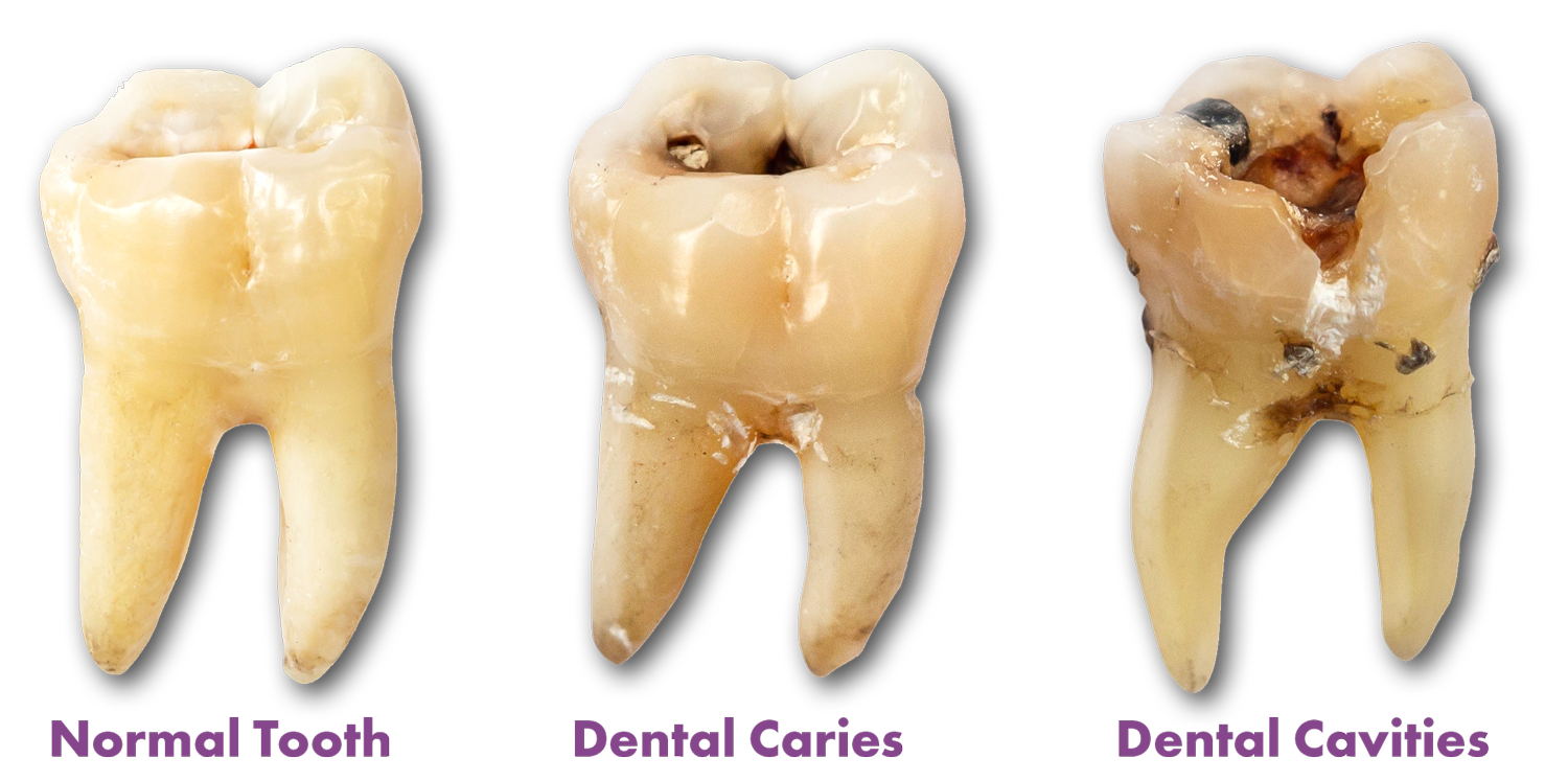
- Clinical Appearance: Clinically, a cavity can range from a small, well-defined pit to a larger, irregular hole. The color can vary from off-white to brown or black, depending on factors like diet, stain accumulation, and the stage of decay. The texture will be soft and irregular, easily picked up by a dental explorer.
- Radiographic Detection: On radiographs, active cavities appear as distinct radiolucent areas. The size and depth of the radiolucency correspond to the extent of tooth structure lost. Advanced cavities can expose the underlying dentin, which is more radiolucent than enamel, making the lesion appear larger on an X-ray. CBCT can precisely map the 3D extent of these cavities, even those hidden beneath restorations.
- Fluorescence and EIS: These technologies will show increasingly pronounced deviations from healthy tissue readings as the lesion progresses, indicating significant mineral loss and structural compromise.
Hidden Caries and Complex Lesions
Technology is crucial for identifying caries that are not easily visible clinically.
- Under Restorations: Decay can form beneath fillings or crowns, termed “secondary caries.” Digital radiography is essential for detecting these, as the radiolucency will appear around the margins of the restoration. CBCT offers even greater detail in assessing the extent and depth of such hidden decay.
- Root Caries: In patients with gum recession, the root surfaces of teeth become exposed and are highly susceptible to decay. These lesions often appear as soft, leathery areas that are dark in color. Digital bitewings can sometimes detect the initial stages, but close clinical examination and tactile assessment are critical. Advanced imaging might be needed if the extent is unclear.

The Future of Caries Visualization and Management
The technological trajectory in dentistry points towards increasingly sophisticated methods for not only detecting caries but also for predicting risk and personalizing treatment. The visualization of dental caries is becoming more precise, objective, and predictive, moving us closer to a truly preventative model of oral healthcare. As these technologies become more accessible and integrated into routine dental practice, our understanding of “what do dental caries look like” will continue to evolve, empowering both clinicians and patients with better tools for maintaining optimal oral health.
aViewFromTheCave is a participant in the Amazon Services LLC Associates Program, an affiliate advertising program designed to provide a means for sites to earn advertising fees by advertising and linking to Amazon.com. Amazon, the Amazon logo, AmazonSupply, and the AmazonSupply logo are trademarks of Amazon.com, Inc. or its affiliates. As an Amazon Associate we earn affiliate commissions from qualifying purchases.