A Dual-energy X-ray Absorptiometry (DXA) scan, often referred to as a DEXA scan, is a non-invasive and remarkably precise imaging technique primarily utilized to measure bone mineral density (BMD). While its most common application is in diagnosing osteoporosis and assessing fracture risk, the advanced capabilities of DXA technology extend to body composition analysis, making it a valuable tool across various health and wellness domains. In essence, a DXA scan provides a detailed, quantitative insight into the internal structure of the body, going beyond what is visible to the naked eye or discernible through simpler diagnostic methods. This technology has become a cornerstone in preventative healthcare and performance optimization due to its accuracy, safety, and the actionable data it yields.
The Science Behind DXA: Unpacking the Technology
At its core, DXA technology leverages the fundamental principle of differential absorption of X-rays by different tissues. The “dual-energy” aspect is key to its diagnostic prowess. The machine emits two beams of X-rays, each at a distinct energy level. These beams pass through the body, and as they do, they are absorbed to varying degrees by different tissues – predominantly bone, soft tissue (muscle and fat), and lean mass. Specialized detectors beneath the patient measure the amount of X-ray energy that has passed through.
How the Dual-Energy Principle Works
The difference in how these two X-ray beams are absorbed allows the DXA scanner to differentiate between bone mineral content and soft tissue mass. Bones, with their higher mineral content, absorb more X-rays than soft tissues. By analyzing the attenuation (reduction in intensity) of each X-ray beam, the sophisticated software within the DXA machine can mathematically separate and quantify these different tissue types.
- Low-Energy Beam: This beam is more readily absorbed by bone mineral.
- High-Energy Beam: This beam penetrates more deeply and is absorbed by both bone and soft tissue.
The software compares the attenuation of the two beams, effectively “subtracting” the soft tissue absorption from the total absorption to isolate and measure the bone mineral density. This process is repeated for specific regions of interest (ROIs) within the body, most commonly the hip and spine for osteoporosis assessment, but also the forearm and whole body for broader analyses.
Scan Procedure and Patient Experience
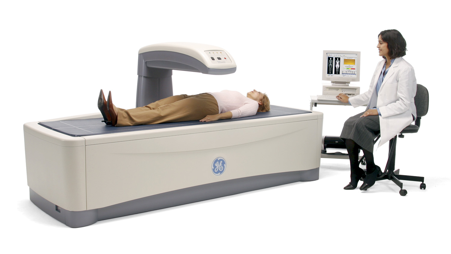
The DXA scanning procedure is straightforward and relatively quick, typically lasting between 10 to 30 minutes depending on the areas being scanned. The patient lies comfortably on a padded table while a C-arm-like scanner arm moves slowly over their body, emitting the X-ray beams. It is crucial for the patient to remain still during the scan to ensure the accuracy of the images and data. There is no injection or contrast material required, and the radiation dose is very low, comparable to that of a standard X-ray. Patients can usually resume their normal activities immediately after the scan. The resulting data is then processed into detailed reports, often including visual representations of the scanned areas.
Applications of DXA Scans: Beyond Bone Health
While DXA’s primary role has historically been in evaluating bone health, its sophisticated body composition analysis capabilities have broadened its applicability significantly. This dual functionality makes DXA a versatile tool for healthcare professionals and individuals seeking a comprehensive understanding of their physical makeup.
Bone Mineral Density (BMD) Assessment
This is the most well-known application of DXA scans. By measuring the BMD at key skeletal sites like the lumbar spine, proximal femur (hip), and sometimes the forearm, DXA scans can:
- Diagnose Osteoporosis: This condition, characterized by low bone mass and structural deterioration, significantly increases fracture risk. DXA scans provide T-scores and Z-scores that compare an individual’s BMD to that of a healthy young adult and a person of the same age and sex, respectively.
- Assess Fracture Risk: Even in the absence of full-blown osteoporosis, low BMD can indicate an increased risk of fractures, particularly in postmenopausal women, older men, and individuals with certain medical conditions or on specific medications.
- Monitor Treatment Efficacy: For individuals undergoing treatment for osteoporosis, DXA scans are used to track the effectiveness of medications and lifestyle interventions by measuring changes in BMD over time.
- Detect Osteopenia: This is a condition where bone density is lower than normal but not yet severe enough to be classified as osteoporosis. DXA helps identify this intermediate stage, allowing for early intervention.
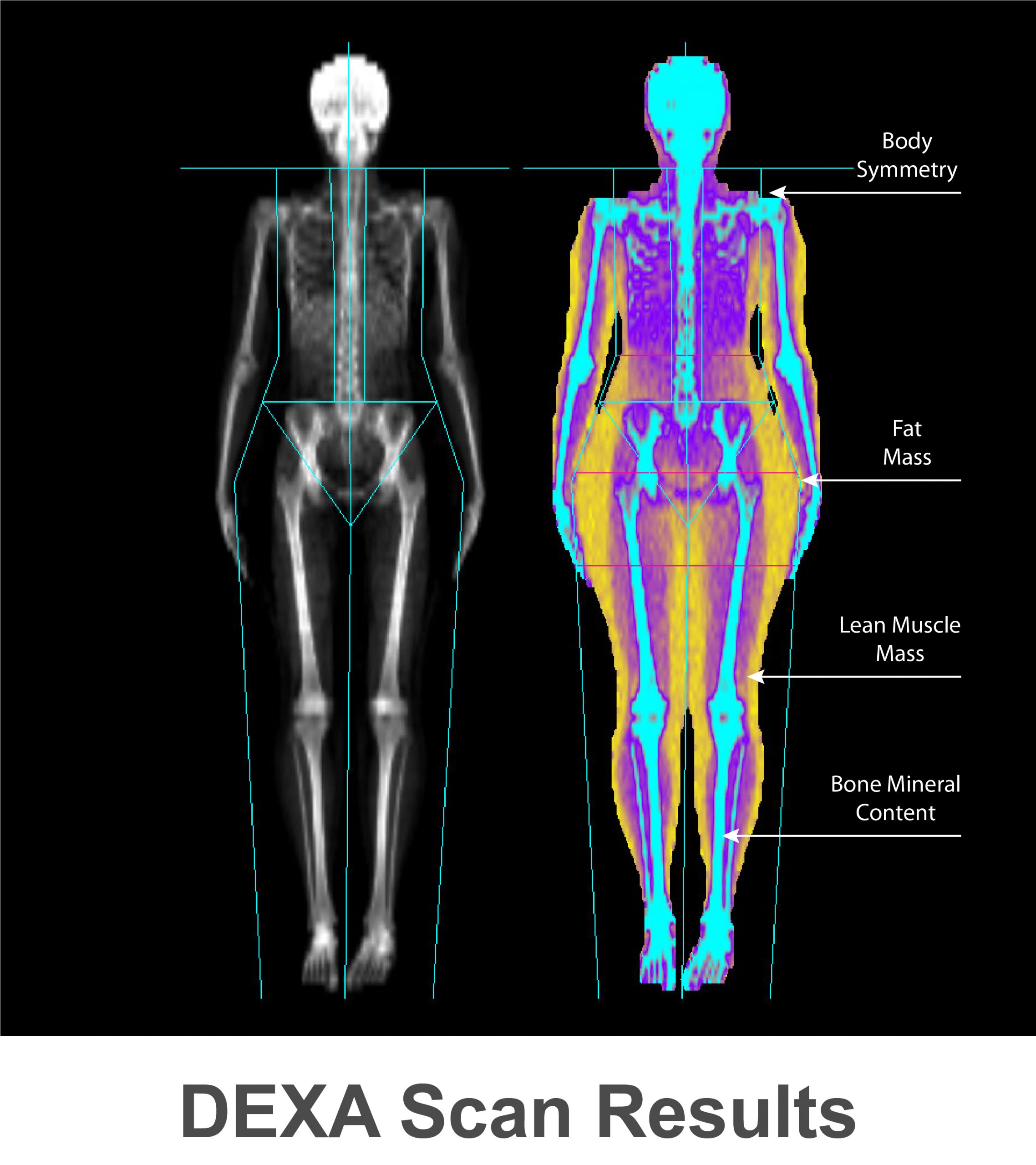
Body Composition Analysis
Beyond bones, DXA scans provide highly accurate measurements of the body’s different tissue components. This analysis includes:
- Lean Mass: This encompasses muscle mass and organ tissue. Tracking lean mass is crucial for understanding muscle health, athletic performance, and recovery from illness or injury.
- Fat Mass: This quantifies the total amount of fat in the body, which can be further analyzed for regional distribution (e.g., android vs. gynoid fat). This information is invaluable for weight management, assessing metabolic health, and identifying risks associated with abdominal obesity.
- Bone Mass: While already covered in BMD assessment, it’s also a component of the overall body composition breakdown.
- Percent Body Fat: DXA offers a precise measurement of an individual’s percentage of body fat, which is a more robust indicator of health risks than simple BMI alone.
- Visceral Fat: Some advanced DXA systems can also estimate visceral adipose tissue (VAT), the fat surrounding internal organs, which is a significant risk factor for cardiovascular disease and type 2 diabetes.
This detailed body composition data is particularly useful for:

- Athletes and Fitness Enthusiasts: Optimizing training programs, monitoring muscle gain or loss, and understanding body composition changes in relation to performance.
- Weight Management Programs: Providing objective data to track progress beyond just scale weight, focusing on fat loss and muscle preservation.
- Nutritional Guidance: Tailoring dietary plans based on an individual’s specific body composition needs.
- Sarcopenia Assessment: Identifying age-related muscle loss, a critical factor in mobility and overall health in older adults.
- Disease Monitoring: Tracking changes in body composition associated with chronic diseases like HIV, cancer, and kidney disease.
Understanding DXA Scan Results: T-Scores, Z-Scores, and Body Composition Metrics
Interpreting the data generated by a DXA scan is a collaborative effort between the patient and their healthcare provider. The reports are designed to be informative, but understanding the key metrics is essential for making informed decisions about health and treatment.
Bone Density Metrics: T-Scores and Z-Scores
The primary metrics for assessing bone health from a DXA scan are T-scores and Z-scores. These scores are derived by comparing the patient’s BMD to reference populations:
- T-Score: This compares the patient’s BMD to the average BMD of a healthy 30-year-old adult of the same sex.
- T-score of -1.0 or higher: Normal bone density.
- T-score between -1.0 and -2.5: Osteopenia (low bone mass).
- T-score of -2.5 or lower: Osteoporosis.
- Z-Score: This compares the patient’s BMD to the average BMD of individuals of the same age, sex, and ethnicity. Z-scores are particularly useful for children, premenopausal women, and younger men.
- Z-score of -2.0 or lower: May indicate an abnormal bone density for the individual’s age and sex, prompting further investigation into secondary causes of bone loss.
Body Composition Metrics Explained
The body composition analysis provides a breakdown of lean mass, fat mass, and bone mass. Key metrics include:
- Total Fat Mass (kg or lbs): The absolute weight of fat in the body.
- Total Lean Mass (kg or lbs): The absolute weight of muscle, organs, and other non-fat tissue.
- Percent Body Fat (%): The proportion of total body weight that is fat. This is often considered a more informative metric than total fat mass alone.
- Appendicular Lean Mass (ALM): The lean mass of the arms and legs. This is a key indicator for sarcopenia (age-related muscle loss).
- Regional Fat Distribution: Some reports may break down fat mass by region (e.g., trunk, arms, legs, android/gynoid regions) to identify areas of particular concern, such as excess visceral fat.
The interpretation of these metrics is highly individualized and depends on the patient’s age, sex, activity level, health status, and goals. A healthcare provider will review these scores in conjunction with other clinical information to provide a comprehensive assessment.
Advantages, Limitations, and Future of DXA Technology
DXA technology has revolutionized bone health assessment and body composition analysis, offering a highly accurate and accessible means of understanding the body’s internal composition. However, like any diagnostic tool, it has its strengths and weaknesses, and ongoing advancements promise even greater utility.
Strengths of DXA Scans
- High Accuracy and Precision: DXA is considered the gold standard for measuring bone mineral density, offering precise and reproducible results. Its body composition analysis is also significantly more accurate than many other methods.
- Low Radiation Exposure: The radiation dose is minimal, making it safe for repeat scans, which is crucial for monitoring treatment progress.
- Non-Invasive: The procedure is painless and requires no injections or special preparation, making it well-tolerated by most patients.
- Comprehensive Data: DXA provides a wealth of information about bone health and body composition, allowing for a holistic assessment of an individual’s health status.
- Versatility: Applicable across a wide range of demographics, from young athletes to the elderly, and for various health conditions.
Limitations of DXA Scans
- Limited Resolution for Subtle Fractures: While excellent for measuring BMD, DXA is not ideal for detecting small or hairline fractures. Other imaging techniques like CT scans or MRIs may be needed for such diagnoses.
- Artifacts from Calcifications or Aortic Calcifications: Dense calcifications in soft tissues can sometimes interfere with BMD measurements, leading to artificially high readings.
- Body Size Limitations: Patients with extreme obesity may exceed the weight or size limits of some DXA tables and scanners, preventing a complete scan.
- Cost and Accessibility: While becoming more widespread, DXA scanners are expensive, which can limit their availability in some regions or healthcare settings.
- Interpretation Dependency: The accuracy of the interpretation relies heavily on the skill of the technologist performing the scan and the radiologist or physician interpreting the results.
The Evolving Landscape of DXA Technology
The field of DXA technology is continuously evolving. Future advancements are likely to focus on:
- Improved Image Resolution: Enhancements in detector technology and software algorithms could lead to even finer detail in bone structure and soft tissue analysis.
- Faster Scan Times: Innovations in hardware and data processing may reduce scan duration, further improving patient comfort and throughput.
- AI Integration: Artificial intelligence and machine learning are poised to play a significant role in automating image analysis, identifying subtle patterns, and providing more personalized risk assessments. This could lead to earlier and more accurate diagnoses.
- Enhanced Body Composition Analysis: More detailed breakdown of tissue types, including precise measurements of visceral fat, muscle fiber type, and hydration levels, could become more commonplace.
- Integration with Other Data Sources: Future DXA systems might integrate data from wearable devices, genetic information, and other health records to provide a more comprehensive and predictive health profile.
In conclusion, the DXA scan is a powerful and multifaceted diagnostic tool that goes far beyond simply assessing bone health. Its precise measurement of bone mineral density and detailed body composition analysis make it an indispensable technology in modern healthcare, sports science, and wellness. As technology continues to advance, the DXA scan is set to become even more integral to personalized health strategies and proactive disease management.
aViewFromTheCave is a participant in the Amazon Services LLC Associates Program, an affiliate advertising program designed to provide a means for sites to earn advertising fees by advertising and linking to Amazon.com. Amazon, the Amazon logo, AmazonSupply, and the AmazonSupply logo are trademarks of Amazon.com, Inc. or its affiliates. As an Amazon Associate we earn affiliate commissions from qualifying purchases.