
The term “barium enema” might sound archaic to some, conjuring images of outdated medical practices. However, this diagnostic imaging technique, while having evolved considerably, remains a vital tool in understanding the health of the lower gastrointestinal tract. From a technological standpoint, a barium enema represents a sophisticated application of X-ray technology, contrast agents, and precise imaging methodologies to visualize and diagnose conditions that might otherwise remain hidden. It’s a testament to how fundamental imaging principles, when combined with advanced technological execution, can offer profound insights into human anatomy and pathology. This article delves into the technological underpinnings, the evolution of its implementation, and the modern role of the barium enema within the broader landscape of medical imaging and diagnostic tools.
The Technological Foundation: How Barium Enema Works
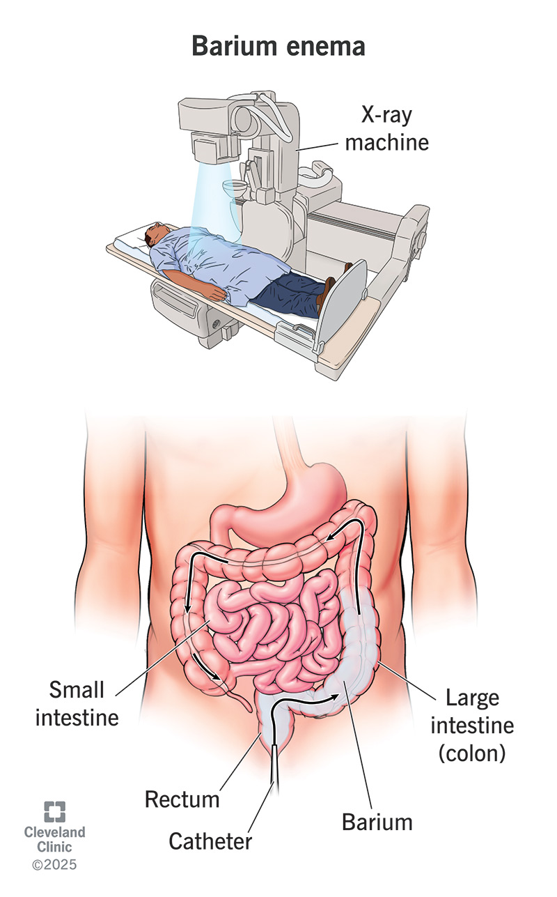
At its core, a barium enema is a radiological examination. This means it leverages the principles of X-rays to create images of internal body structures. However, X-rays alone are not always sufficient for visualizing soft tissues like the colon. This is where the technological ingenuity of the barium enema truly shines, through the use of a radiopaque contrast agent and precise imaging techniques.
The Role of Barium Sulfate: A Radiopaque Marvel
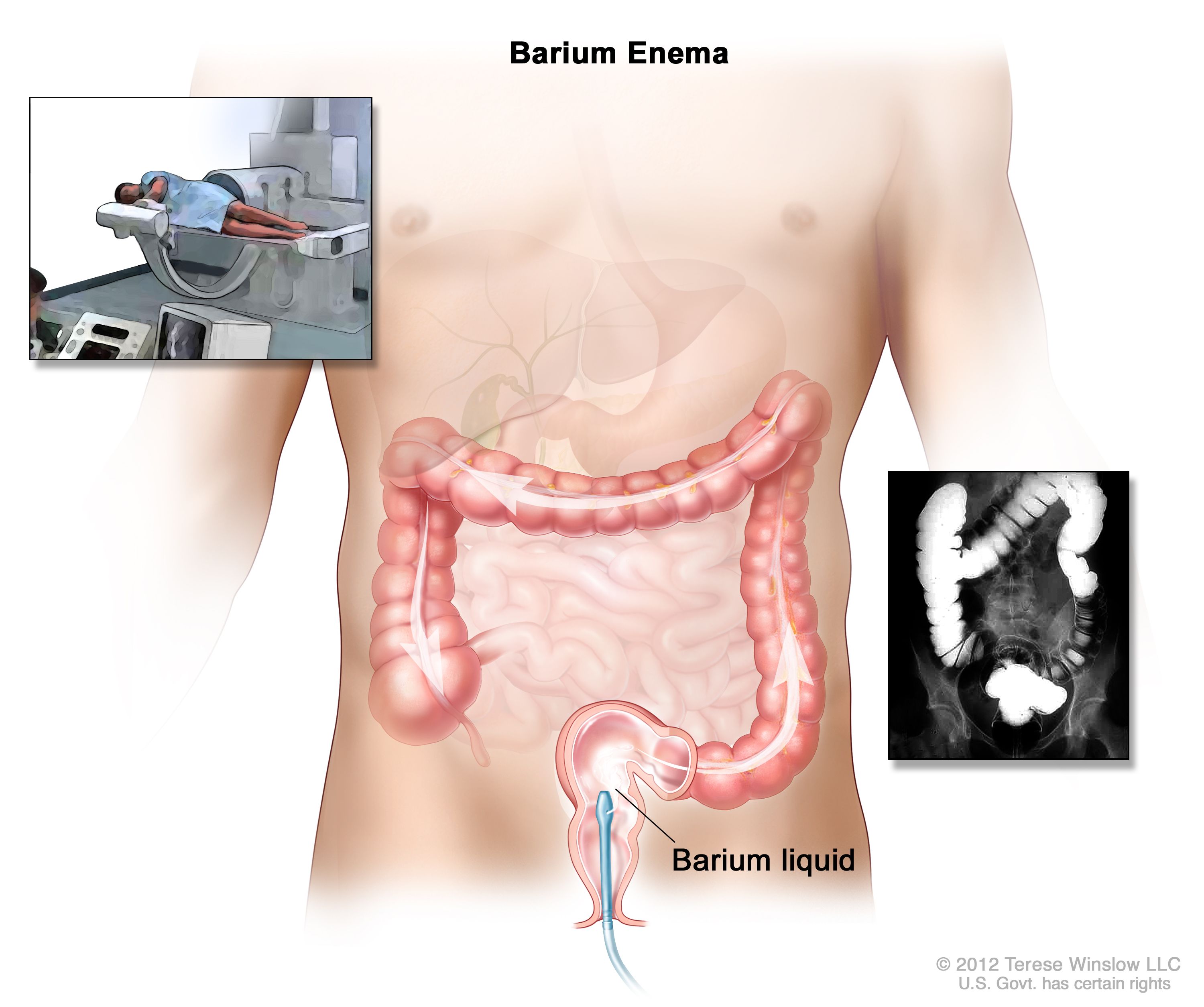
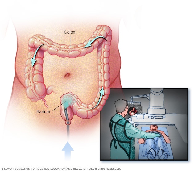
The “barium” in barium enema refers to barium sulfate, a chemical compound with a unique property: it is radiopaque. This means it absorbs X-rays much more effectively than surrounding tissues. When introduced into the colon, barium sulfate coats the inner lining of the organ, effectively outlining its shape and structure. This contrast agent is prepared as a liquid suspension, often described as a chalky fluid. The precise formulation and consistency of the barium sulfate suspension are critical technological considerations, ensuring optimal coating without causing undue discomfort or distortion to the colonic walls. Technological advancements in the production of barium sulfate have led to suspensions that are smoother, easier to administer, and less prone to sedimentation, thereby enhancing the quality of the diagnostic images.
X-ray Imaging and Fluoroscopy: Capturing the Dynamic Image
The diagnostic power of the barium enema is realized through the application of X-ray technology. After the barium sulfate has been introduced, X-rays are passed through the abdomen. These rays are differentially absorbed by the body’s tissues. The barium sulfate, being highly absorbent, appears white on the resulting X-ray image, while air within the colon (if used in double-contrast studies) appears black, and the soft tissues of the bowel wall and surrounding organs appear in shades of gray.
More advanced than simple static X-rays, fluoroscopy is a key technological component of the barium enema procedure. Fluoroscopy allows for real-time, dynamic visualization of the barium as it moves through the colon. This is achieved by a continuous or pulsed X-ray beam projected onto a fluorescent screen or, more commonly today, onto a digital detector. The radiologist can observe the barium coating the colon, identify any areas where the coating is disrupted, and assess the motility and contour of the bowel as it is manipulated. This dynamic imaging capability is crucial for identifying subtle abnormalities such as polyps, diverticula, strictures, or areas of inflammation that might be missed on static images. The technological evolution from early fluorescent screens to modern digital fluoroscopy, with its improved image resolution, lower radiation doses, and digital recording capabilities, has significantly enhanced the diagnostic accuracy and safety of the barium enema.
Double-Contrast Technique: Enhancing Visualization
A significant technological advancement in barium enema procedures is the double-contrast technique. While a single-contrast barium enema primarily visualizes the mucosal surface as a single layer, the double-contrast method employs both barium sulfate and air (or sometimes carbon dioxide) to create a more detailed and three-dimensional image. After the barium is instilled, a controlled amount of air is pumped into the colon, inflating it. This inflation separates the barium coating from the bowel wall, allowing for a clearer visualization of the mucosal lining. The air acts as a negative contrast agent, appearing black on the X-ray image, while the barium remains white. This creates a stark contrast between the barium-coated surface and the air-filled lumen, highlighting subtle irregularities, polyps, and other lesions with greater precision. The technological control over the inflation pressure and the rate of air insufflation is paramount to the success of this technique, ensuring patient comfort and optimal image quality.
Evolution of the Technology: From Early Radiography to Digital Precision
The journey of the barium enema mirrors the broader technological advancements in medical imaging. What began as a rudimentary application of X-rays has been refined through decades of innovation, leading to safer, more effective, and more detailed diagnostic procedures.
Early Applications and Limitations
The early days of radiology saw the advent of barium studies in the early 20th century. These procedures were groundbreaking, offering the first non-invasive glimpses into the digestive tract. However, early X-ray equipment was bulky, and image quality was often limited. The barium suspensions were also less refined, leading to inconsistencies in coating and potential for patient discomfort. Radiation doses were also considerably higher than what is considered acceptable today. Despite these limitations, the barium enema proved invaluable in diagnosing conditions like colonic obstruction, tumors, and inflammatory bowel diseases, paving the way for future diagnostic imaging techniques.
The Digital Revolution: Enhancing Image Quality and Safety
The most significant technological leap for barium enema, and indeed for all of diagnostic imaging, has been the digital revolution. Traditional X-ray film has largely been replaced by digital radiography and fluoroscopy systems. In a digital system, the X-ray beam strikes a digital detector, which converts the radiation into an electronic signal. This signal is then processed by a computer to create a digital image. The advantages of this technology are manifold:
- Improved Image Resolution: Digital detectors can capture finer details, leading to more precise visualization of the colonic mucosa.
- Reduced Radiation Dose: Digital systems are often more sensitive to X-rays, allowing for lower radiation doses to be used while maintaining excellent image quality. This is a critical safety advancement.
- Post-Processing Capabilities: Digital images can be manipulated on a computer. Radiologists can adjust brightness, contrast, and zoom in on specific areas of interest without re-exposing the patient to radiation. This enhances diagnostic interpretation.
- Archiving and Sharing: Digital images can be easily stored electronically, shared with other medical professionals, and accessed for follow-up examinations, streamlining patient care and collaboration.
- PACS Integration: These digital images are typically integrated into Picture Archiving and Communication Systems (PACS), allowing for seamless retrieval and management of patient imaging data within healthcare institutions.
The development of advanced imaging processing algorithms further refines these digital images, sharpening edges, reducing noise, and enhancing the visibility of subtle mucosal changes.
Modern Applications and Technological Integration
Despite the rise of other advanced imaging modalities like CT colonography and colonoscopy, the barium enema retains a specific niche in modern diagnostic medicine, largely due to its technological advantages in certain clinical scenarios.
Barium Enema in Specific Clinical Contexts
While colonoscopy is often considered the gold standard for visualizing the entire colon and performing biopsies, the barium enema offers distinct advantages in certain situations. For instance, in cases of suspected bowel perforation, where the risk of insufflating air into the peritoneal cavity during colonoscopy is high, a single-contrast barium enema might be preferred. The controlled instillation of barium, with its lower risk of leakage, can provide essential information about the location and extent of the perforation.
Furthermore, for patients who are unable to undergo a full colonoscopy due to contraindications like severe heart disease, respiratory illness, or recent bowel surgery, a barium enema can serve as a valuable alternative for screening and diagnostic purposes. Its ability to visualize the overall structure and contour of the colon makes it effective in detecting large polyps, strictures, or masses.
The technological precision of double-contrast barium enema is particularly adept at delineating the extent of inflammatory bowel disease, such as ulcerative colitis or Crohn’s disease, by clearly showing the irregular mucosal surface and any narrowing of the bowel lumen. It can also be instrumental in diagnosing diverticular disease and identifying the presence of fistulas or abscesses in complicated cases.
The Role of Advanced Imaging Techniques and AI
While the fundamental principles of barium enema remain, ongoing technological research explores further enhancements. This includes investigating new types of contrast agents that might offer improved visualization or reduced risk of complications. The integration of artificial intelligence (AI) is also a growing area of interest within diagnostic imaging. While not yet widespread for barium enemas, AI algorithms are being developed to assist in the interpretation of medical images. These algorithms could potentially:
- Automate Lesion Detection: AI could be trained to identify suspicious areas on barium enema images, such as polyps or masses, flagging them for radiologist review.
- Quantify Disease: AI could assist in measuring the extent of colonic wall thickening or the degree of lumen narrowing, providing objective data for disease management.
- Improve Workflow Efficiency: By pre-analyzing images or highlighting areas of concern, AI could help radiologists interpret studies more quickly and accurately.
These technological integrations promise to further optimize the diagnostic yield and efficiency of barium enema examinations, ensuring its continued relevance in the modern medical landscape. The barium enema, therefore, is not merely a historical artifact but a dynamic diagnostic tool that has adapted and evolved with technological progress, continuing to offer critical insights into colonic health.
aViewFromTheCave is a participant in the Amazon Services LLC Associates Program, an affiliate advertising program designed to provide a means for sites to earn advertising fees by advertising and linking to Amazon.com. Amazon, the Amazon logo, AmazonSupply, and the AmazonSupply logo are trademarks of Amazon.com, Inc. or its affiliates. As an Amazon Associate we earn affiliate commissions from qualifying purchases.