
The human ear, a marvel of biological engineering, is responsible for our sense of hearing and balance. It’s a complex structure, and like any intricate system, it can be susceptible to disruptions. Among the most common ailments affecting the ear is the ear infection, medically known as otitis. While we often feel the discomfort and pain associated with these infections, understanding what they look like internally can provide valuable insight into the disease process and the importance of timely medical intervention. This article delves into the visual manifestations of ear infections as observed during medical examinations, offering a glimpse into the hidden world within our auditory canals and middle ears.
Understanding the Anatomy of the Ear: A Prerequisite to Visualizing Infection
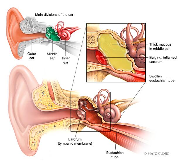
Before we can appreciate the visual cues of an ear infection, a foundational understanding of the ear’s anatomy is crucial. The ear is broadly divided into three main sections: the outer ear, the middle ear, and the inner ear. Each plays a distinct role in the process of hearing and maintaining balance, and each can be the site of an infection.
The Outer Ear: The First Line of Defense
The outer ear comprises the auricle (the visible part of the ear) and the external auditory canal, also known as the ear canal. This canal is a narrow tube that leads from the auricle to the eardrum (tympanic membrane). Its primary functions are to collect sound waves and channel them towards the eardrum, and to protect the delicate structures further inside. The skin lining the ear canal contains glands that produce cerumen, or earwax, which has protective and lubricating properties.
- Visual Characteristics of a Healthy Outer Ear: In a healthy state, the ear canal appears clear and somewhat moist, with a light pink or pale red hue. The presence of a small amount of earwax, typically yellowish or brownish, is normal and protective. The eardrum, viewed through an otoscope at the end of the canal, should be pearly gray, translucent, and exhibit landmarks like the malleus (a small bone). The cone of light, a reflection of the otoscope light, should be visible.
The Middle Ear: The Vibrational Hub
Behind the eardrum lies the middle ear, an air-filled cavity. This space houses the three smallest bones in the human body: the malleus, incus, and stapes, collectively known as the ossicles. These ossicles transmit vibrations from the eardrum to the inner ear. The middle ear is connected to the nasopharynx (the back of the nose and upper throat) by the Eustachian tube. This tube is vital for equalizing pressure in the middle ear and draining any fluid that may accumulate.
- The Role of the Eustachian Tube in Infection: The Eustachian tube is a common pathway for bacteria and viruses to enter the middle ear, particularly in children whose tubes are shorter and more horizontally oriented. Inflammation or blockage of this tube, often due to colds or allergies, can lead to fluid buildup in the middle ear, creating a breeding ground for infection.
The Inner Ear: The Gateway to the Brain
The innermost part of the ear is the inner ear, which contains the cochlea (responsible for hearing) and the semicircular canals (responsible for balance). These structures are filled with fluid and contain sensory cells that convert mechanical vibrations into electrical signals, which are then transmitted to the brain via the auditory nerve. Infections in the inner ear (labyrinthitis) are less common but can lead to more severe symptoms like dizziness and hearing loss.
Visualizing Otitis Externa: Infection of the Outer Ear
Otitis externa, commonly known as “swimmer’s ear,” is an infection of the external auditory canal. It often arises from prolonged exposure to water, which can strip away protective earwax, making the skin more vulnerable to bacterial or fungal invasion. Minor trauma, such as scratching the ear canal with a cotton swab or fingernail, can also initiate the infection.
What a Doctor Sees During Otoscopy
When a physician examines a patient with suspected otitis externa using an otoscope, the visual findings can be striking.
- Inflammation and Redness: The most prominent sign is significant inflammation of the ear canal’s lining. The pale pink color of a healthy canal will be replaced by a bright, angry red. This erythema can range from mild to severe, often making the entire canal appear swollen and inflamed.
- Swelling and Edema: The tissues within the ear canal become edematous, meaning they are swollen due to fluid accumulation. This swelling can narrow the ear canal considerably, making it difficult to visualize the eardrum. In severe cases, the canal might be almost completely occluded by swollen tissue.
- Discharge (Otorrhea): Infected ear canals often produce discharge. This can vary in appearance depending on the type of pathogen involved.
- Bacterial Infections: Typically result in a thick, purulent discharge that can be yellow, green, or even white. It may have a foul odor.
- Fungal Infections (Otomycosis): Often present with a white, cheesy, or cotton-like discharge, sometimes accompanied by black or brown flecks if the fungus is Aspergillus.
- Painful to the Touch: The inflammation and swelling make the ear canal exquisitely tender. Even the slightest touch to the auricle or gentle manipulation of the ear canal can elicit intense pain for the patient.
- Debris and Crusts: Dead skin cells, inflammatory exudate, and the discharge itself can form crusts and debris within the ear canal, further obscuring visualization of deeper structures.
Visualizing Otitis Media: Infection of the Middle Ear
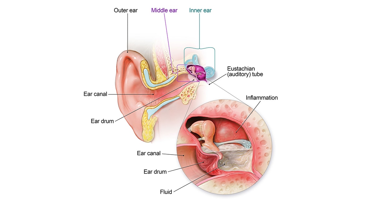
Otitis media is far more common, especially in children, and typically follows an upper respiratory infection. When bacteria or viruses ascend the Eustachian tube, they can infect the fluid that may have accumulated in the middle ear, leading to pus formation.
Acute Otitis Media (AOM): The Classic Picture
Acute otitis media is a sudden onset infection characterized by inflammation and the presence of fluid in the middle ear.
- The Eardrum’s Appearance: During otoscopic examination, the eardrum is the key indicator. In AOM, the eardrum loses its normal pearly gray, translucent appearance. Instead, it often appears:
- Red and Inflamed (Hyperemic): The redness is not just superficial; the entire eardrum can be intensely red, indicating increased blood flow due to inflammation.
- Bulging: The accumulation of pus and pressure behind the eardrum forces it outward, causing it to bulge significantly. This bulging is a hallmark of AOM and is often accompanied by severe pain as the pressure increases.
- Opaque and Loss of Landmarks: The normal translucency is lost, making it difficult or impossible to see the ossicles or the cone of light. The overall color can range from pink to a milky white or even yellowish if pus is visible through the membrane.
- Decreased Mobility: When a pneumatic otoscope is used, which gently blows a puff of air into the ear canal, the eardrum of a healthy ear will move. In AOM, the inflamed and fluid-filled eardrum will show significantly reduced or absent mobility due to the pressure and effusion.
- Absence of Discharge (Usually): In uncomplicated AOM, there is typically no discharge visible in the ear canal because the infection is confined to the middle ear. However, if the infection is severe enough, the eardrum can rupture, leading to pus draining into the ear canal.
- Perforation of the Eardrum: In cases of severe pressure and infection, the eardrum may perforate (tear). When this happens, pus or infected fluid drains out of the middle ear into the ear canal. Visually, this would be seen as a hole or tear in the eardrum, with purulent discharge flowing from it. The color of the discharge is typically thick, yellowish, or greenish.
Otitis Media with Effusion (OME): The “Silent” Infection
Otitis media with effusion, also known as “glue ear,” is characterized by the presence of fluid in the middle ear without signs of acute infection. This fluid can be thin and watery or thick and viscous (“glue-like”). While not acutely painful, OME can lead to hearing loss and speech development delays, especially in children.
- Eardrum Appearance in OME:
- Retracted or Neutral Position: Unlike AOM, the eardrum in OME is often retracted inward or is in a neutral position, not bulging.
- Dull and Opaque: The eardrum appears dull and opaque, losing its pearly gray luster.
- Fluid Levels or Bubbles: The most distinctive visual sign is the presence of fluid behind the eardrum. This may be seen as a fluid line (meniscus) or as air bubbles trapped within the viscous fluid. These are often best visualized with a pneumatic otoscope or by using a special otoscope attachment called a video otoscope which allows for magnification and detailed imaging.
- Loss of Mobility: Similar to AOM, the eardrum in OME has significantly reduced mobility on pneumatic otoscopy due to the presence of the effusion.
Visualizing Otitis Interna: Rarer but More Serious
Infections of the inner ear (labyrinthitis) are less common and are often a complication of untreated middle ear infections or can arise from viral infections spreading from the brain.
Challenges in Direct Visualization
Unlike the outer and middle ear, the inner ear structures are not directly visible through an otoscope. Diagnosis of labyrinthitis is typically based on a constellation of symptoms, including severe dizziness (vertigo), nausea, vomiting, and hearing loss, rather than direct visual inspection of the infected area.
Indirect Signs and Imaging
While direct visualization is impossible, medical professionals may infer inner ear involvement based on the overall condition of the ear. If a severe middle ear infection (otitis media) is present and has been left untreated, it can spread to the inner ear. Imaging techniques like MRI or CT scans may be used to assess the extent of inflammation or damage to the inner ear structures, though these are typically reserved for complex or persistent cases.
The Importance of Visual Diagnosis and Medical Intervention
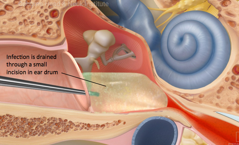
The ability to visually identify the signs of ear infections inside the ear is fundamental to accurate diagnosis and effective treatment. An otoscope, a simple yet powerful medical instrument, allows healthcare providers to peer into the ear canal and assess the condition of the eardrum.
- Differentiating Infection Types: The distinct visual characteristics of otitis externa, acute otitis media, and otitis media with effusion guide the clinician in choosing the appropriate treatment. For example, the red, bulging eardrum of AOM typically requires antibiotics, while the dull, fluid-filled eardrum of OME might be managed with watchful waiting or other interventions.
- Assessing Severity: The degree of redness, swelling, bulging, or presence of discharge helps gauge the severity of the infection. This information is critical for determining the urgency of treatment and the potential for complications.
- Monitoring Treatment Efficacy: Follow-up examinations allow doctors to visually monitor the resolution of inflammation, the disappearance of fluid, and the return of the eardrum to its normal appearance, confirming that the treatment is working.
- Preventing Complications: Early and accurate visual diagnosis facilitates prompt treatment, which is crucial for preventing complications such as chronic ear infections, hearing loss, or the spread of infection to other parts of the body.
In conclusion, while the internal structures of the ear are hidden from plain view, medical professionals utilize specialized tools and their expertise to visualize the tell-tale signs of infection. From the inflamed, weeping canal of otitis externa to the red, bulging eardrum of acute otitis media, these visual cues are invaluable in understanding, diagnosing, and effectively treating these common yet potentially impactful conditions. The seemingly simple act of looking inside the ear with an otoscope unlocks a world of diagnostic information, underscoring the power of visual inspection in modern medicine.
aViewFromTheCave is a participant in the Amazon Services LLC Associates Program, an affiliate advertising program designed to provide a means for sites to earn advertising fees by advertising and linking to Amazon.com. Amazon, the Amazon logo, AmazonSupply, and the AmazonSupply logo are trademarks of Amazon.com, Inc. or its affiliates. As an Amazon Associate we earn affiliate commissions from qualifying purchases.