The presence of intestinal worms in stool is a visible indicator of parasitic infection, a condition that affects millions globally. While historically identified through manual inspection, modern technology has revolutionized the diagnosis and management of these often-debilitating ailments. This article delves into the technological advancements that enable us to accurately identify and understand the visual characteristics of worms in feces, moving beyond simple observation to sophisticated diagnostic methodologies.
The Visual Spectrum of Intestinal Worms in Feces
The visual identification of intestinal worms in stool can range from the readily apparent to the subtly disguised. Understanding these varied appearances is the first step in any diagnostic process, and technology now plays a crucial role in enhancing our ability to perceive these differences.
Macroscopic Identification: What to Look For
When one speaks of “worms in poo,” they often refer to the macroscopic, or visible, forms of intestinal parasites. These are typically adult or larval stages of helminths (worms) that have been expelled from the host’s body. The appearance can vary significantly based on the specific type of worm.
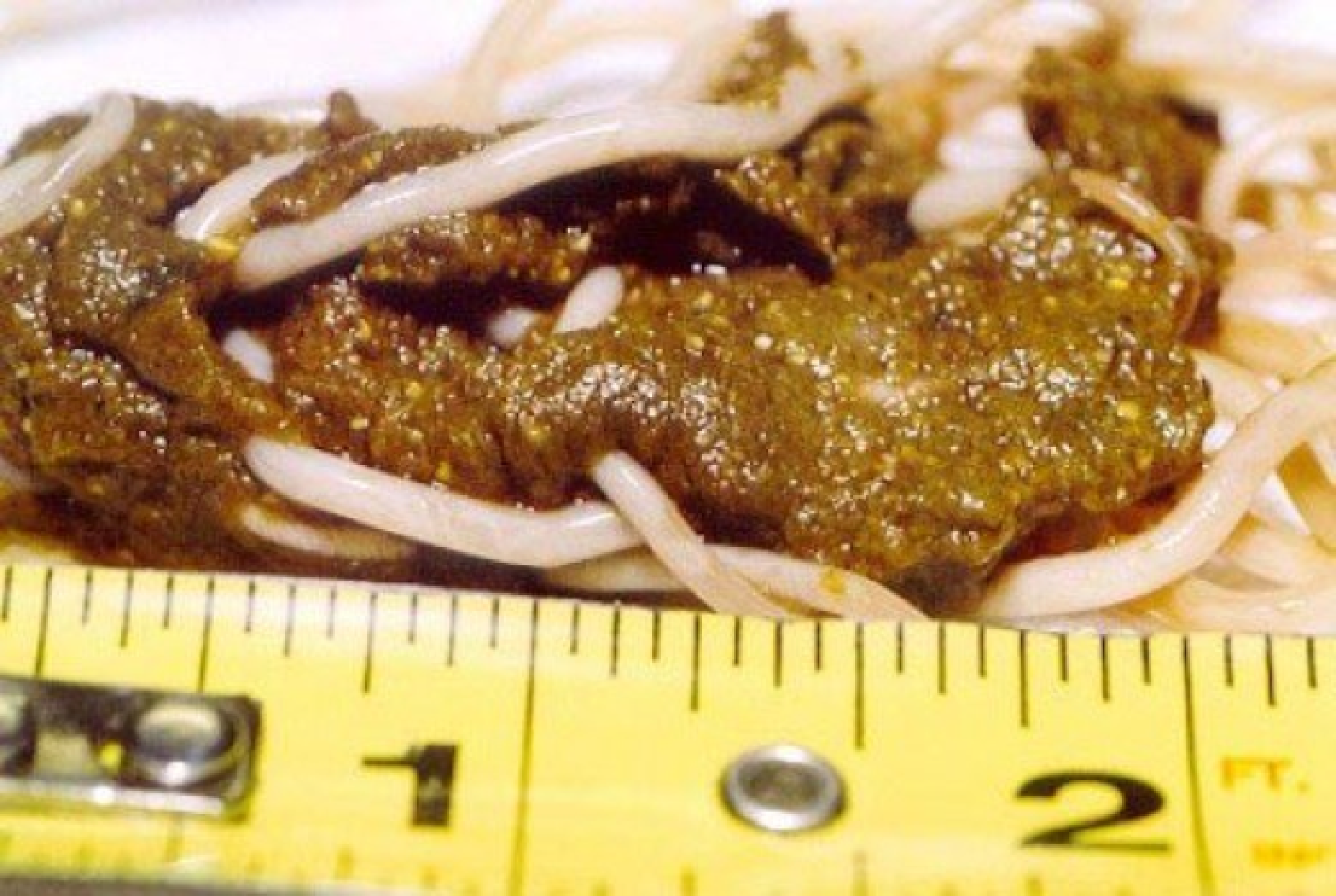
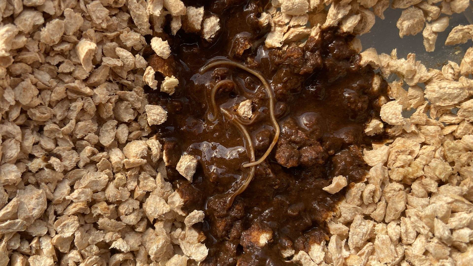
Roundworms (Ascaris lumbricoides)
- Appearance: These are among the largest intestinal roundworms, often resembling earthworms. They are typically pinkish-white or cream-colored and can range from 15 to 35 centimeters in length.
- In Stool: Whole, live roundworms are less common to see unless the infection is heavy. More frequently, fragments of worms, or tangled masses of worms, might be observed. Their texture is firm and cylindrical.
- Technological Aid: While direct visual identification is primary, microscopy can confirm the presence of eggs, which are much smaller and require magnification. Digital imaging software can be used to analyze the morphology of observed worm segments for more precise identification in clinical settings.
Pinworms (Enterobius vermicularis)
- Appearance: Pinworms are small, white, thread-like nematodes. Adult females are about 8–13 mm long, and males are smaller, about 2–5 mm.
- In Stool: Seeing whole pinworms in stool is rare because they typically lay their eggs on the perianal skin, not in the feces. However, in cases of severe infestation, they might be found. Their small size and white color can make them difficult to distinguish from mucus strands or undigested food particles.
- Technological Aid: The gold standard for pinworm diagnosis remains the “tape test,” where a piece of clear tape is pressed against the perianal area. This tape is then examined under a microscope. While not directly observing worms in poo, this technology leverages microscopic imaging for detection. Advanced digital microscopes with high-resolution cameras can capture detailed images of eggs and even adult pinworms for remote analysis and record-keeping.
Tapeworms (Cestodes)
- Appearance: Tapeworms are segmented, ribbon-like parasites. The segments are called proglottids.
- In Stool: Proglottids are the most common visual evidence of tapeworms in feces. They may appear as small, white, rice-grain-like segments that can move independently when fresh. Longer segments, or even entire tapeworms (though rare), might be expelled.
- Technological Aid: Microscopic examination of proglottids and eggs is crucial for species identification. Digital calipers and imaging analysis software can measure the dimensions of proglottids and identify specific morphological features, aiding in distinguishing between different tapeworm species. Advanced imaging techniques, such as confocal microscopy, can provide detailed cross-sections of proglottids for precise anatomical analysis.
Hookworms (Ancylostoma duodenale and Necator americanus)
- Appearance: Adult hookworms are small (1–2 cm long), slender, and have a hook-like anterior end.
- In Stool: Adult hookworms are rarely seen in stool as they attach to the intestinal wall. Diagnosis is typically made by identifying their eggs under a microscope.
- Technological Aid: Microscopic examination of stool samples for hookworm eggs is the standard. Advanced hematology analyzers and automated microscopy systems can process large numbers of samples and identify microscopic structures with high accuracy. AI-powered image recognition algorithms are being developed to automatically detect and classify parasitic eggs in stool samples, significantly speeding up the diagnostic process.
Microscopic Clues: Eggs and Larvae
Beyond visible worms, the eggs and larval stages of many intestinal parasites are microscopic, requiring specialized technological tools for their detection and identification.
Egg Morphology
- Varied Shapes and Sizes: Parasitic eggs come in a vast array of shapes, sizes, and surface textures. Some are oval, others spherical; some are smooth, while others have thick, spiny, or pitted shells. These characteristics are critical for accurate identification of the parasite species.
- Technological Aid: Microscopy is fundamental. High-powered compound microscopes are essential for visualizing these minute structures. Digital microscopes with integrated cameras allow for real-time image capture, measurement, and digital archiving. This facilitates sharing of images for consultation with experts and for educational purposes. Automated stool analysis systems utilize image processing and pattern recognition algorithms to scan slides for eggs, flagging potential positives for human review. This technology drastically reduces the time required for manual examination and minimizes human error.
Larval Stages
- Active Forms: Some parasites, such as Strongyloides stercoralis, can exist in larval forms in stool. These larvae are motile and can be identified under the microscope.
- Technological Aid: Phase-contrast microscopy is particularly useful for visualizing the motility and detailed structures of larvae, which might be obscured by their transparency under standard bright-field microscopy. Digital video recording of microscopic observations allows for detailed analysis of larval movement patterns, which can aid in species identification.
Technological Innovations in Parasite Detection
The detection of intestinal worms has moved far beyond the simple visual inspection of stool. A suite of advanced technologies now supports clinicians and researchers in identifying and characterizing these infections.
Advanced Microscopy and Imaging
The core of parasitic diagnosis lies in microscopic examination, and technology has continually pushed the boundaries of what is possible.
High-Resolution Digital Microscopy
- Enhanced Visualization: Modern digital microscopes offer superior resolution and magnification compared to traditional optical microscopes. They provide crystal-clear images of parasitic eggs and larvae, allowing for more detailed morphological analysis.
- Image Capture and Analysis: Integrated high-resolution cameras enable the capture of digital images and videos, which can be stored, shared, and analyzed using specialized software. This facilitates remote diagnostics, telemedicine applications, and the creation of comprehensive digital patient records.
- Quantitative Analysis: Software can perform measurements of egg size and shape, count parasite forms, and even assist in identifying species based on learned patterns, improving accuracy and efficiency.
Fluorescence Microscopy
- Specific Staining: Certain stains can bind specifically to parasitic structures, making them more visible and distinct under fluorescence microscopy. This can enhance the detection of difficult-to-see parasites or their components.
- In Situ Hybridization (ISH): While more common in research, ISH techniques combined with fluorescent probes can detect specific nucleic acid sequences of parasites within host cells or stool samples, offering a high degree of specificity.
Molecular Diagnostic Techniques
The advent of molecular biology has introduced highly sensitive and specific methods for detecting parasites, often identifying them at their genetic level.
Polymerase Chain Reaction (PCR)
- DNA Amplification: PCR techniques amplify specific DNA sequences of parasites, allowing for their detection even when only minute amounts of genetic material are present. This is invaluable for identifying infections where eggs or adult worms are scarce or absent in stool.
- Species-Specific Detection: By designing primers that target unique DNA regions of different parasite species, PCR can differentiate between closely related organisms that might appear morphologically similar under a microscope.
- Quantitative PCR (qPCR): This variation of PCR can also quantify the parasite load in a sample, providing an estimate of infection intensity, which can be useful for monitoring treatment efficacy.
Next-Generation Sequencing (NGS)
- Metagenomic Analysis: NGS allows for the simultaneous identification of DNA from all organisms present in a sample, including a broad spectrum of parasites. This metagenomic approach can uncover coinfections or identify novel parasitic species.
- High Throughput: NGS platforms can process large numbers of samples, making them suitable for epidemiological studies and public health surveillance.
Automation and Artificial Intelligence (AI) in Diagnostics
The sheer volume of stool samples processed in clinical laboratories has driven the development of automated systems and AI-powered solutions.
Automated Stool Analyzers
- High-Throughput Screening: These systems use robotics and advanced imaging to automatically prepare, scan, and analyze stool samples for the presence of parasites, significantly increasing throughput and reducing manual labor.
- Standardized Protocols: Automation ensures standardized sample preparation and analysis, leading to greater consistency and reproducibility of results.
AI-Powered Image Recognition
- Pattern Recognition: Machine learning algorithms are trained on vast datasets of microscopic images to recognize the characteristic shapes and features of parasitic eggs and larvae.
- Assisted Diagnosis: AI systems can act as an invaluable second reader for laboratory technicians, highlighting suspicious areas on slides and flagging potential positives, thus improving diagnostic accuracy and speed.
- Predictive Analytics: As AI evolves, it may also contribute to predicting infection risk or identifying patterns in diagnostic data that could inform public health interventions.
The Role of Digital Health Platforms and Telemedicine
The integration of diagnostic technologies with digital platforms is transforming how parasitic infections are managed, particularly in remote or underserved areas.
Telemedicine for Parasitic Diagnosis
- Remote Consultation: Patients can submit images or videos of stool samples (where appropriate and safe) to healthcare providers via secure digital platforms. This allows for preliminary assessment and guidance without the need for an immediate in-person visit.
- Expert Interpretation: Digital images of microscopic slides can be shared with parasitology experts located anywhere in the world for rapid consultation and definitive diagnosis. This is particularly beneficial in areas with limited local expertise.
Mobile Health (mHealth) Applications
- Patient Education: Apps can provide users with information about parasitic infections, including visual guides to what worms or eggs might look like in stool, and advice on when to seek medical attention.
- Symptom Tracking and Reporting: mHealth tools can help individuals track symptoms, record potential exposures, and report findings, which can be valuable data for both individual patient care and public health surveillance.

Data Management and Public Health Surveillance
- Digital Databases: Electronic health records and specialized laboratory information systems allow for the secure storage and analysis of diagnostic data.
- Trend Analysis: Aggregated data from these systems can be used to monitor the prevalence and geographic distribution of parasitic infections, identify outbreak patterns, and inform public health strategies and interventions.
- AI for Epidemiology: AI algorithms can analyze large epidemiological datasets to identify risk factors, predict disease spread, and optimize resource allocation for control programs.
In conclusion, while the initial question about the appearance of worms in poo might seem rudimentary, the technological advancements in its diagnosis reveal a sophisticated and evolving field. From high-resolution digital microscopy and molecular diagnostics to AI-powered analysis and telemedicine, technology is continuously enhancing our ability to detect, identify, and ultimately combat intestinal worm infections, improving global health outcomes.
aViewFromTheCave is a participant in the Amazon Services LLC Associates Program, an affiliate advertising program designed to provide a means for sites to earn advertising fees by advertising and linking to Amazon.com. Amazon, the Amazon logo, AmazonSupply, and the AmazonSupply logo are trademarks of Amazon.com, Inc. or its affiliates. As an Amazon Associate we earn affiliate commissions from qualifying purchases.