In the rapidly evolving landscape of health technology, the traditional method of peering through a manual microscope to identify pathogens is being replaced by sophisticated digital imaging, artificial intelligence (AI), and automated laboratory systems. When we ask the question, “What does Candida look like in urine?” from a technological perspective, we are no longer just describing a biological entity. We are discussing data points, pixel recognition, and the algorithmic interpretation of fungal morphology.
The digital transformation of clinical diagnostics has turned the identification of Candida albicans and other fungal species into a masterclass in high-resolution imaging and machine learning. This article explores the cutting-edge technology used to visualize, identify, and quantify Candida in urinary samples, highlighting how software and hardware are revolutionizing the field of medical microbiology.
The Evolution of Visualization: From Manual Optics to Digital Imaging
The journey of identifying fungal elements in urine begins with the hardware. Traditionally, a laboratory technician would prepare a slide of urine sediment and manually scan for budding yeast cells or pseudohyphae. Today, this process has been digitized and automated through high-throughput urinalysis platforms.
From Manual Microscopy to Automated Digital Urinalysis (ADU)
Modern clinical laboratories now utilize Automated Digital Urinalysis (ADU) systems. These machines are marvels of optical engineering. Instead of a static slide, the urine sample flows through a specialized chamber where a high-speed digital camera captures thousands of images per minute. These systems use “flow-cell” technology, ensuring that every particle in the urine—including Candida—is photographed in high definition. For a technologist, Candida is no longer just a speck on a lens; it is a high-resolution JPEG or TIFF file ready for algorithmic analysis.
High-Resolution Imaging and Phase-Contrast Technology
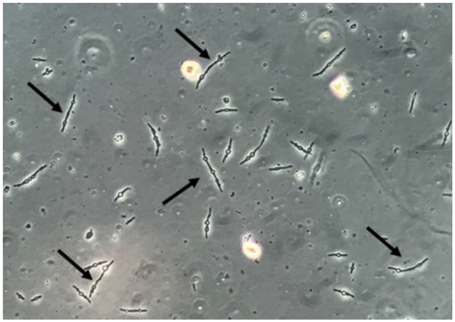
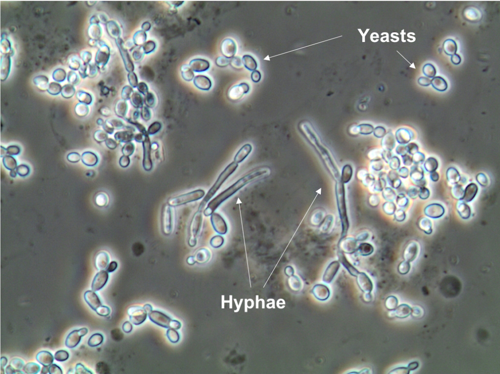
To see what Candida “looks like” digitally, technology utilizes phase-contrast microscopy integrated into digital sensors. Candida cells are often translucent, making them difficult to see under standard light. Phase-contrast tech converts phase shifts in light passing through the transparent specimen into brightness changes in the image. This results in high-contrast digital renderings where the oval shapes of yeast cells and the elongated structures of pseudohyphae stand out against the complex background of the urine sediment, which may contain crystals, bacteria, and epithelial cells.
AI and Machine Learning in Fungal Identification
The true “tech” breakthrough in answering what Candida looks like in urine lies in the software. Identifying a yeast cell among millions of other particles is a task perfectly suited for Artificial Intelligence, specifically Computer Vision.
Deep Learning and Convolutional Neural Networks (CNNs)
Modern diagnostic software employs Convolutional Neural Networks (CNNs) to identify Candida. These AI models are trained on massive datasets containing millions of labeled images of urinary particles. When the digital camera captures a frame, the software breaks the image down into layers, analyzing edges, shapes, and textures.
- Budding Yeast Identification: The AI looks for specific geometric ratios—the “mother-daughter” cell relationship characterized by a smaller oval protruding from a larger one.
- Pseudohyphae Detection: The software identifies long, tube-like structures with distinct indentations.
By utilizing deep learning, these tools can distinguish between Candida and similar-looking objects, such as red blood cells or calcium oxalate crystals, with a level of precision that often exceeds human capability.
Reducing “Noise” through Algorithmic Filtering
Urine is a “noisy” environment for a sensor. It contains various debris that can lead to false positives. Advanced software suites use noise-reduction algorithms to clear the digital field of view. By applying filters that ignore background artifacts and focus on the refractive index of fungal cell walls, the technology provides a “clean” visualization of the Candida presence. This process, known as image segmentation, allows the software to isolate the fungal elements from the rest of the sample for closer inspection.
Telemedicine and the Rise of At-Home Diagnostic Tech
The question of what Candida looks like in urine is moving from the hospital lab into the palm of the consumer’s hand. The “MedTech” sector is seeing a surge in smartphone-integrated diagnostic tools designed for home use.
Smartphone-Integrated Urinalysis Kits
Startups are currently developing kits that utilize the high-powered cameras already present in modern smartphones. These kits often involve a dipstick or a small collection device that interfaces with a mobile app. Using the phone’s macro lens and a specialized light-attachment tool, the app can capture images of the sample.
The “look” of Candida in this context is interpreted by an on-device AI. The app processes the image locally or sends it to a secure cloud server, where it compares the visual data against a database of fungal morphologies. Within minutes, the user receives a digital report—a far cry from waiting days for a traditional culture.
Cloud-Based Data Sharing and Security
In the tech-driven diagnostic model, the visual data of Candida is not just an image; it is a secure medical record. Telemedicine platforms integrate these diagnostic images into Electronic Health Records (EHR). This involves sophisticated data pipelines that must comply with HIPAA (Health Insurance Portability and Accountability Act) and GDPR (General Data Protection Regulation). The “look” of the fungus is encrypted and transmitted to healthcare providers, allowing for remote “over-read” where a human pathologist verifies the AI’s findings via a high-bandwidth digital interface.
Big Data and Predictive Analytics in Mycology
When technology visualizes Candida across thousands of patients simultaneously, it creates a “Big Data” opportunity. What Candida looks like in urine at a macro level can inform public health trends.
Tracking Fungal Resistance through Data Aggregation
Large diagnostic networks aggregate the digital results of urinalysis to track the prevalence of certain Candida species. If the software identifies an uptick in Candida auris—a highly resistant and dangerous strain—the data can be visualized on a geographic heatmap. This allows tech-driven health organizations to predict outbreaks and monitor the efficacy of antifungal treatments in real-time. The visual identification of the fungus thus becomes a tool for predictive modeling.
Blockchain for Diagnostic Integrity
As we move toward a more decentralized diagnostic framework, ensuring the integrity of the “image” is paramount. Some tech firms are exploring the use of blockchain to create an immutable ledger for diagnostic images. When an AI identifies Candida in a urine sample, the metadata—timestamp, device ID, and the visual hash—is recorded on a blockchain. This prevents data tampering and ensures that the diagnostic “look” of the sample remains consistent from the moment of capture to the moment of treatment.
Future Trends: Nanotechnology and Real-Time Bio-Sensors
The future of seeing what Candida looks like in urine goes beyond pixels and enters the realm of molecular sensors and nanotechnology.
Nano-Sensors and Lab-on-a-Chip
We are approaching an era of “Lab-on-a-Chip” (LoC) technology. These are microfluidic devices that can detect the molecular “signature” of Candida rather than just its visual shape. These chips use gold nanoparticles or carbon nanotubes that change electrical conductivity when they bind to Candida proteins. In this tech paradigm, what Candida “looks like” is a specific electrical signal or a fluorescent glow on a micro-scale sensor.
Integration with Wearable Health Tech
The ultimate goal of health tech is continuous monitoring. Researchers are investigating the possibility of “smart toilets” or wearable sensors that can perform real-time urinalysis. These devices would use spectroscopic sensors to analyze the chemical and biological composition of urine. Instead of a one-time visual check, the “look” of Candida would be a trend line on a user’s smartwatch, alerting them to a fungal overgrowth before physical symptoms even manifest.

Conclusion: The Digital Future of Microbiology
The question “what does candida look like in urine” has been fundamentally redefined by technology. It is no longer a simple description of a biological organism; it is a complex intersection of high-speed digital imaging, deep-learning algorithms, and secure data transmission.
From the high-tech laboratories using AI-driven convolutional neural networks to the smartphone apps bringing diagnostics to the home, technology has made the visualization of Candida faster, more accurate, and more accessible. As we look forward to nanotechnology and real-time bio-sensors, the “visual” aspect of diagnostics will continue to evolve, turning the invisible world of fungal pathogens into actionable, digital intelligence. In this tech-driven era, seeing is no longer just believing—it is data-driven certainty.
aViewFromTheCave is a participant in the Amazon Services LLC Associates Program, an affiliate advertising program designed to provide a means for sites to earn advertising fees by advertising and linking to Amazon.com. Amazon, the Amazon logo, AmazonSupply, and the AmazonSupply logo are trademarks of Amazon.com, Inc. or its affiliates. As an Amazon Associate we earn affiliate commissions from qualifying purchases.