The intersection of medical science and cutting-edge engineering has birthed one of the most sophisticated diagnostic tools in history: the Cardiac Magnetic Resonance (CMR) scan. While the general public often views an MRI as a standard medical procedure, from a tech perspective, it is a masterclass in high-performance computing, signal processing, and quantum physics. When we ask “what does a heart MRI show,” we aren’t just looking at a picture of a muscle; we are looking at a complex data visualization of biological mechanics, fluid dynamics, and tissue composition.
As technology continues to evolve, the resolution and depth of what a heart MRI can reveal have expanded exponentially. Today, heart MRI tech is the gold standard for non-invasive cardiac assessment, providing insights that were previously only possible through invasive biopsies or surgeries.
The Engineering Marvel: How Magnetic Resonance Visualizes the Heart
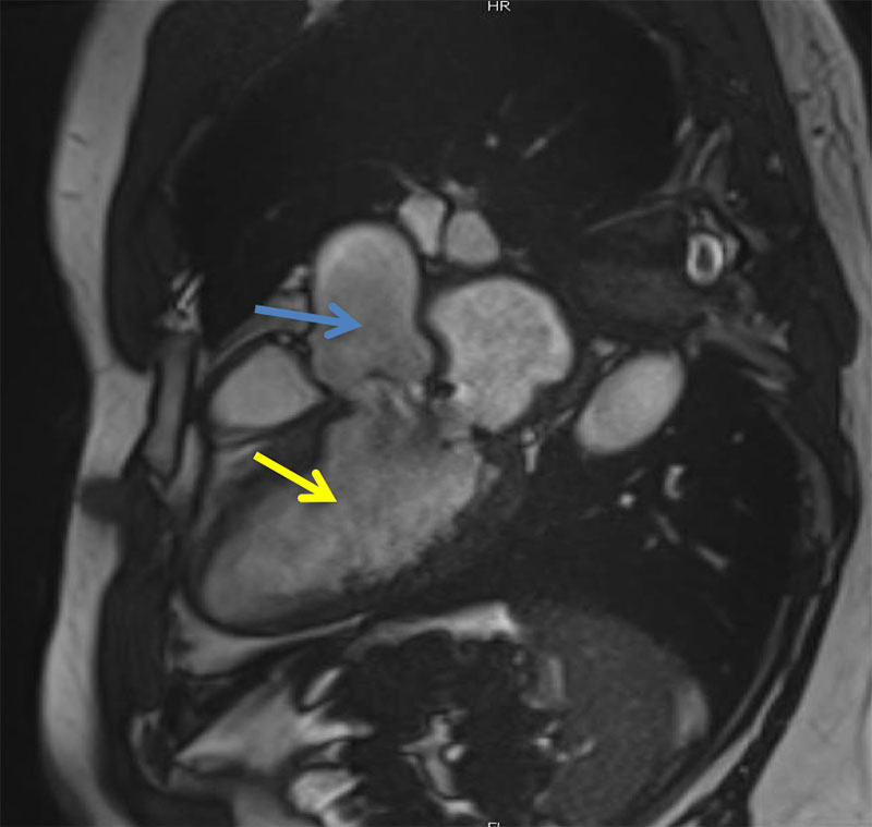
At its core, a heart MRI is a feat of hardware engineering. Unlike X-rays or CT scans that use ionizing radiation, an MRI utilizes powerful magnetic fields and radiofrequency (RF) pulses to manipulate the spin of hydrogen protons in the body. In the context of the heart—a constantly moving organ—this technology requires incredible precision to capture clear data.
High-Field Strength Magnets and Signal Processing
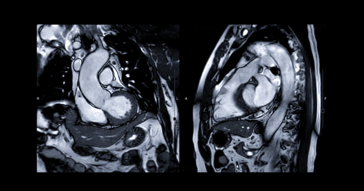
The quality of what a heart MRI “shows” is directly tied to the Tesla (T) rating of the magnet. Modern clinical settings typically use 1.5T or 3T scanners. The higher the field strength, the better the signal-to-noise ratio, allowing for higher-resolution images. To capture a beating heart, the tech must synchronize with the patient’s ECG (electrocardiogram). This “gating” technology ensures that data is collected at the exact same point in the cardiac cycle, preventing motion blur and allowing the software to reconstruct a crisp, moving image of the heart’s valves and chambers.
Multi-Parametric Mapping: Beyond Basic Images
One of the most significant technological leaps in CMR is “mapping.” Standard MRI images are weighted (T1 or T2) to show anatomy, but parametric mapping allows the tech to show the actual physical properties of the heart tissue. T1 mapping, for example, can quantify the amount of interstitial fibrosis (microscopic scarring) within the heart muscle. This shift from qualitative “looking at a picture” to quantitative “measuring tissue values” is a massive trend in medical technology, turning the MRI into a digital laboratory.
Software-Driven Diagnostics: Analyzing Cardiac Function and Structure
The hardware gathers the data, but the software is what interprets it into something a human can understand. The modern suite of cardiac imaging software transforms raw signals into detailed 3D and 4D models. When we look at what a heart MRI shows, we are seeing the result of complex algorithms processing terabytes of information.
Cine Imaging and Real-Time Motion Tracking
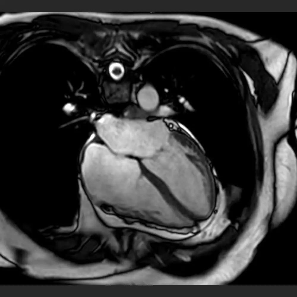
What a heart MRI shows most vividly is the heart in motion. Through “Cine” sequences, the software creates a movie-like loop of the heart beating. Tech advancements now allow for “feature tracking,” where software follows specific points on the heart wall to measure “strain.” Strain imaging is a high-tech way of seeing how much the heart muscle deforms during a contraction. This can reveal subtle dysfunction long before the heart’s overall pumping capacity (ejection fraction) starts to drop.
Tissue Characterization: Identifying Fibrosis and Edema
A heart MRI is unique because of its ability to perform “Late Gadolinium Enhancement” (LGE). By injecting a gadolinium-based contrast agent, the tech can show exactly where heart tissue has been damaged. Gadolinium washes out of healthy tissue quickly but lingers in scarred or infarcted (dead) tissue. On the screen, this “shows” up as bright white spots against a dark background of healthy muscle. This software-enabled visualization is critical for distinguishing between different types of cardiomyopathies or identifying the remnants of a silent heart attack.
The AI Revolution in Cardiac MRI Interpretation
We are currently in a transition period where Artificial Intelligence (AI) and Machine Learning (ML) are taking over the heavy lifting of MRI interpretation. The sheer volume of data produced by a single heart MRI can be overwhelming for human radiologists, but it is the perfect playground for AI tools.
Automated Segmentation and Measurement
In the past, a technician would have to manually “trace” the outlines of the heart’s chambers across dozens of image slices to calculate volumes. Today, AI-driven software uses deep learning models to perform automated segmentation in seconds. This technology not only saves time but also removes human variability. What the MRI “shows” is now more accurate because the measurements are standardized by algorithms trained on millions of previous scans.
Predictive Analytics: Turning Images into Prognoses
The next frontier of heart MRI tech is predictive analytics. By feeding CMR data into neural networks, researchers are developing tools that can predict a patient’s risk of future heart failure or arrhythmias. The MRI doesn’t just show what the heart looks like now; it provides the data necessary for AI to model what the heart might do in five years. This shift from diagnostic imaging to predictive modeling is a hallmark of the modern “Digital Twin” trend in health tech.
Digital Security and Data Integration in Modern Radiology
As heart MRIs become more data-intensive, the infrastructure surrounding them must become more robust. A single CMR study can produce hundreds of high-resolution images, creating a significant challenge for storage, transmission, and security.
DICOM Standards and Cloud-Based Image Sharing
The “Digital Imaging and Communications in Medicine” (DICOM) standard is the backbone of how MRI data is stored and shared. However, the tech trend is moving toward cloud-based PACS (Picture Archiving and Communication Systems). This allows specialists across the globe to view a heart MRI in real-time. The “show” is no longer confined to a single hospital terminal; it is a mobile, accessible digital asset that can be integrated into a patient’s comprehensive Electronic Health Record (EHR).
Protecting Sensitive Biometric Data
With the rise of AI and cloud sharing, digital security has become a primary concern. A heart MRI contains highly sensitive biometric information. Modern MRI tech must incorporate end-to-end encryption and strict access controls to prevent data breaches. As we move toward more personalized “Tech-Health,” ensuring that the 3D map of a person’s heart is as secure as their bank account is a top priority for developers and cybersecurity experts in the medical field.
The Future of Heart Tech: 4D Flow and Virtual Reality
As we look forward, the question of “what does a heart MRI show” will have even more complex answers. One of the most exciting emerging technologies is 4D Flow MRI. This tech allows for the visualization and quantification of blood flow in three dimensions over time (the fourth dimension).
Imagine a “weather map” of the blood moving through your aorta, showing turbulence, pressure changes, and shear stress on the vessel walls. Furthermore, this data is now being exported into Virtual Reality (VR) environments, allowing surgeons to “walk through” a patient’s heart before they ever make an incision. This is the ultimate evolution of the MRI: it is no longer just a scan, but a fully immersive digital reconstruction of human life.
Conclusion
A heart MRI is far more than a medical test; it is the pinnacle of current imaging technology. By leveraging high-field magnetism, sophisticated signal processing, AI-driven analysis, and secure digital infrastructure, it shows us the intricate reality of the human heart in unprecedented detail. As software continues to eat the world, it is also refining our view of the heart, turning every heartbeat into a data point and every scan into a roadmap for longevity. For those in the tech and medical-engineering space, the heart MRI represents the perfect synergy of hardware and software, providing a clear window into the most vital machine we will ever own.
aViewFromTheCave is a participant in the Amazon Services LLC Associates Program, an affiliate advertising program designed to provide a means for sites to earn advertising fees by advertising and linking to Amazon.com. Amazon, the Amazon logo, AmazonSupply, and the AmazonSupply logo are trademarks of Amazon.com, Inc. or its affiliates. As an Amazon Associate we earn affiliate commissions from qualifying purchases.