
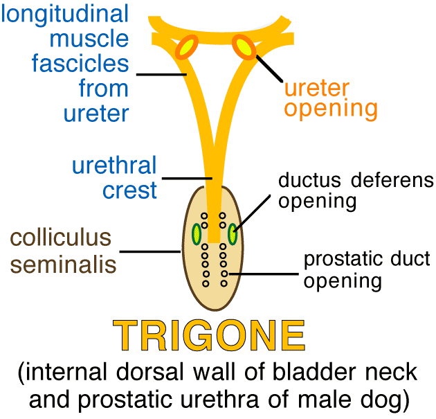
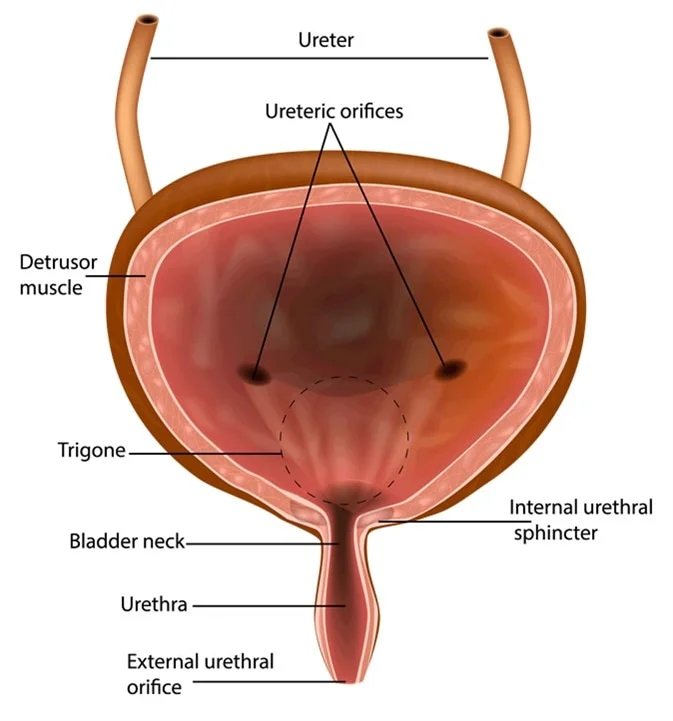
The trigone, a smooth, triangular region located at the base of the urinary bladder, is anatomically distinctive due to its unique embryological origin and crucial functional role in urinary continence and flow. Traditionally, its definition has been purely anatomical: bounded by the two ureteral orifices superiorly and the internal urethral orifice inferiorly. However, in the 21st century, “what defines the trigone” extends far beyond mere static anatomy. It now encompasses a dynamic interplay with cutting-edge technology that enables unprecedented visualization, precise diagnosis, targeted intervention, and sophisticated training. This technological lens provides a deeper, more functional, and clinically actionable definition of the trigone, transforming urological care and research.
Advanced Medical Imaging: Visualizing the Trigone with Unprecedented Clarity
The ability to accurately visualize anatomical structures in vivo has been a cornerstone of medical progress, and the trigone is no exception. Modern medical imaging technologies provide a non-invasive or minimally invasive means to define the trigone’s morphology, assess its integrity, and detect pathologies with remarkable precision, effectively giving us a dynamic, multi-dimensional “definition.”
High-Resolution Ultrasound and Doppler Imaging
Ultrasound remains a first-line diagnostic tool due to its non-invasiveness, real-time capabilities, and cost-effectiveness. High-resolution transabdominal, transrectal, and especially transvaginal or transperineal ultrasound can provide detailed views of the bladder trigone, its muscular layers, and the ureteral jets. Doppler imaging further enhances this by visualizing blood flow, assessing vascularity, and identifying potential obstructions or anomalies in the ureters as they enter the bladder. Advances in 3D/4D ultrasound technology allow for multi-planar reconstruction and dynamic assessment of the trigone’s shape and function during bladder filling and voiding, offering a functional definition that was previously only available through invasive procedures. This real-time feedback loop is crucial for diagnosing conditions like vesicoureteral reflux (VUR) or bladder outlet obstruction.
MRI and CT Scans: Multi-Dimensional Reconstruction
Magnetic Resonance Imaging (MRI) and Computed Tomography (CT) scans offer superior soft tissue contrast and spatial resolution compared to ultrasound, providing a comprehensive anatomical definition of the trigone and surrounding structures. MRI, in particular, excels at delineating soft tissue details without ionizing radiation, making it invaluable for evaluating congenital anomalies, detecting subtle tumors, or assessing inflammatory conditions affecting the trigone. Advanced MRI sequences can map muscular architecture and detect fibrosis, offering insights into the trigone’s structural integrity. CT scans, while involving radiation, provide excellent bony detail and are often used in conjunction with contrast agents to evaluate the entire urinary tract for stones, hydronephrosis, or tumor invasion that might impact the trigone. Sophisticated software tools enable radiologists to reconstruct 3D models of the trigone from these scans, allowing for virtual exploration and precise surgical planning.
Endoscopic and Cystoscopic Technologies
For direct visualization, endoscopic technologies, particularly cystoscopy, remain the gold standard. Modern cystoscopes are sleek, flexible instruments equipped with high-definition cameras, LED illumination, and often multiple working channels. These technological advancements provide urologists with a crisp, magnified view of the trigone’s mucosal surface, allowing for direct assessment of color, texture, vascular patterns, and the orifices of the ureters and urethra. Digital cystoscopy allows for image capture and video recording, essential for documentation, teaching, and longitudinal follow-up. Furthermore, narrow band imaging (NBI) and photodynamic diagnosis (PDD) are advanced optical technologies integrated into cystoscopes that enhance the visualization of subtle mucosal changes, like early signs of bladder cancer, which might be missed under standard white light, thus refining the definition of healthy versus pathological trigonal tissue.
The Role of AI and Machine Learning in Trigonal Diagnostics and Research
Artificial Intelligence (AI) and Machine Learning (ML) are rapidly transforming every facet of medicine, offering powerful tools to process vast amounts of data, identify complex patterns, and predict outcomes. For the trigone, AI/ML platforms are beginning to redefine how we diagnose, prognose, and research conditions affecting this critical area.
Predictive Analytics for Bladder Dysfunction
AI algorithms can analyze patient demographic data, lifestyle factors, medical history, and voiding diaries to predict the likelihood of developing certain bladder dysfunctions, including those related to trigonal pathology. For instance, ML models can be trained on large datasets of patients with conditions like overactive bladder (OAB) or interstitial cystitis, identifying subtle correlations that human analysis might miss. These models can help stratify patient risk, optimize treatment selection, and even predict response to therapies, offering a more personalized and predictive “definition” of a patient’s trigone-related health trajectory.
Automated Image Analysis and Anomaly Detection
One of the most promising applications of AI is in medical imaging. Deep learning algorithms can be trained to analyze ultrasound, MRI, and CT images of the bladder trigone to automatically detect abnormalities such as tumors, inflammation, or structural anomalies. AI-powered software can segment the trigone, measure its dimensions, and compare findings to a vast database of normal and pathological cases, flagging suspicious regions for further human review. This not only enhances diagnostic accuracy but also significantly reduces the time required for image interpretation, providing a faster, more consistent, and objectively defined assessment of the trigone’s state.
AI-Driven Drug Discovery and Therapeutic Development
Beyond diagnostics, AI is accelerating research into new therapies for trigone-related conditions. ML algorithms can analyze molecular data, genetic profiles, and drug compound libraries to identify potential therapeutic targets or compounds that might modulate trigonal function. This approach can drastically shorten the drug discovery pipeline, leading to novel treatments for conditions like bladder pain syndrome or neurogenic bladder, where the trigone often plays a central role. By understanding the complex biochemical and cellular pathways through AI, we gain a deeper, molecular-level “definition” of trigonal health and disease.
Robotic-Assisted Surgery: Precision Interventions in the Trigonal Region
Surgical intervention in the delicate trigonal area demands extreme precision and minimal disruption to surrounding vital structures. Robotic-assisted surgery has emerged as a transformative technology, redefining the capabilities of surgeons and the outcomes for patients undergoing procedures involving the trigone.

Enhanced Dexterity and Visualization in Complex Procedures
Robotic surgical systems, such as the da Vinci platform, provide surgeons with enhanced dexterity, tremor filtration, and a magnified, high-definition 3D view of the surgical field. This allows for incredibly fine movements, improved suturing, and dissection around the trigone, a region dense with nerves and blood vessels crucial for bladder function. For procedures like ureteral re-implantation or bladder augmentation, which often involve the trigone, robotic assistance minimizes trauma to surrounding tissues, leading to faster recovery and reduced complications. The robot effectively “defines” a new standard of surgical precision for the trigone.
Minimally Invasive Approaches to Trigone-Related Conditions
By enabling complex procedures through small incisions, robotic surgery aligns with the principles of minimally invasive surgery. This translates to less post-operative pain, reduced blood loss, shorter hospital stays, and a quicker return to normal activities for patients undergoing trigone-related operations. Conditions such as bladder diverticula near the trigone, vesicovaginal fistulas, or even certain types of bladder cancer can now be addressed robotically, offering a less disruptive “definition” of surgical recovery.
Post-Operative Monitoring and Predictive Maintenance of Surgical Tools
Beyond the operating room, technology continues to play a role. Robotic systems collect vast amounts of data on instrument usage, wear and tear, and procedural nuances. This data can be analyzed to predict when instruments might need maintenance or replacement, ensuring optimal performance and safety. Furthermore, integrating smart sensors in surgical tools could provide real-time feedback during trigonal procedures, helping surgeons avoid excessive force or ensuring correct tissue planes, thus offering an ongoing “definition” of surgical quality and safety.
Virtual Reality and Simulation: Revolutionizing Training for Trigonal Procedures
The complexity and critical nature of the trigone necessitate rigorous training for medical professionals. Virtual Reality (VR) and simulation technologies are fundamentally redefining surgical education, offering safe, immersive, and repeatable environments for mastering trigone-related procedures.
Immersive Anatomical Exploration and Procedural Practice
VR platforms can render highly detailed, interactive 3D models of the urinary bladder and its trigone, allowing students and residents to explore the anatomy from any angle. This goes beyond static textbook images, providing a dynamic and experiential “definition” of the trigonal region. Furthermore, VR simulators allow for realistic practice of endoscopic procedures like cystoscopy or even complex robotic surgeries involving the trigone. Trainees can repeatedly perform steps, identify landmarks, and manage complications in a risk-free environment, honing their skills before operating on real patients.
Surgical Simulators for Competency-Based Education
Advanced haptic feedback systems in surgical simulators replicate the tactile sensations of cutting, suturing, and dissecting tissues around the trigone. This provides a truly immersive experience where trainees can develop both cognitive understanding and psychomotor skills. Simulators can track performance metrics, such as instrument path, force applied, and efficiency, providing objective feedback essential for competency-based education. This allows for a measurable “definition” of a surgeon’s proficiency in handling the delicate trigonal anatomy.
Telemedicine and Remote Collaboration for Urological Expertise
VR and augmented reality (AR) also facilitate remote collaboration and telemedicine. Experienced urologists can guide less experienced surgeons through complex trigonal procedures from a different location, overlaying digital instructions or anatomical references onto the trainee’s view. This “tele-mentoring” capability expands access to specialized expertise, particularly in underserved areas, effectively allowing a broader, geographically distributed “definition” of expert surgical guidance for trigone-related issues.
Future Trends and Emerging Technologies: The Next Frontier for Trigonal Health
The evolution of technology continues unabated, promising even more profound ways to define and interact with the trigone in the years to come.
Wearable Tech for Bladder Monitoring
The development of smart wearables and implantable sensors could revolutionize the long-term monitoring of bladder function, including the trigone’s role. Imagine a discreet patch or an internal sensor that continuously tracks bladder filling, pressure, and voiding patterns, transmitting data to a smartphone or cloud for analysis. This real-time, continuous data would provide an unprecedented functional “definition” of bladder and trigone health, identifying subtle changes before they become symptomatic and enabling proactive management of conditions like OAB or neurogenic bladder.
Gene Editing and Regenerative Medicine for Trigonal Repair
Looking further ahead, advancements in gene editing technologies like CRISPR and regenerative medicine hold immense promise. If trigonal dysfunction is linked to specific genetic predispositions or cellular damage, gene editing could potentially correct these at a fundamental level. Similarly, tissue engineering and stem cell therapies could offer ways to regenerate damaged trigonal muscle or nerves, redefining what is possible in terms of repair and restoration of function. This would move beyond merely defining the trigone’s pathology to redefining its biological integrity.
Big Data Integration for Personalized Treatment Pathways
The ultimate frontier involves integrating all these technological advancements – imaging, AI diagnostics, robotic surgical data, wearable sensor data, and genetic information – into a comprehensive big data ecosystem. AI would then analyze this vast, heterogeneous dataset to create highly personalized “digital twins” of each patient’s urinary system, including a detailed, multi-faceted definition of their trigone. This would allow for truly predictive healthcare, where treatment pathways are tailored to individual needs, optimized for efficacy, and adjusted in real-time, ushering in an era of precision urology that continuously redefines our understanding and management of the trigone.
In conclusion, “what defines the trigone of the urinary bladder” has dramatically expanded from a simple anatomical description to a complex technological narrative. Modern innovations are not merely observing the trigone; they are actively shaping our understanding, diagnosing its conditions with precision, enabling delicate surgical interventions, and educating the next generation of medical professionals. As technology continues its relentless march forward, our definition of the trigone will only grow richer, more dynamic, and increasingly personalized, ultimately benefiting countless patients worldwide.
aViewFromTheCave is a participant in the Amazon Services LLC Associates Program, an affiliate advertising program designed to provide a means for sites to earn advertising fees by advertising and linking to Amazon.com. Amazon, the Amazon logo, AmazonSupply, and the AmazonSupply logo are trademarks of Amazon.com, Inc. or its affiliates. As an Amazon Associate we earn affiliate commissions from qualifying purchases.