In the vast landscape of modern medical technology, few diagnostic tools have revolutionized healthcare as profoundly as Computed Tomography (CT) scans and Magnetic Resonance Imaging (MRI) scans. Both are indispensable for peering inside the human body without surgery, offering detailed views that aid in diagnosing countless conditions. Yet, despite their shared goal of non-invasive internal visualization, the underlying technology, operational principles, and diagnostic strengths of CT and MRI are fundamentally distinct. Understanding these differences isn’t just a matter for medical professionals; it offers a fascinating glimpse into the ingenious applications of physics and engineering that save and improve lives daily. This exploration delves into the technological heart of each, dissecting their mechanics, applications, advantages, and limitations, firmly positioning them as pinnacles of diagnostic innovation within the tech sphere.
Unpacking the Core Technology: How They Work
At the heart of the distinction between CT and MRI lies their disparate technological foundations. One harnesses the power of X-rays, while the other employs magnetic fields and radio waves. This fundamental difference dictates everything from the types of images produced to the conditions they are best suited to diagnose.
Computed Tomography (CT): X-rays in 3D
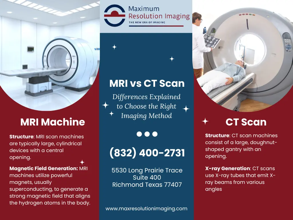
The CT scan, often referred to simply as a CT, is an advanced form of X-ray technology. Instead of producing a single, flat image like a conventional X-ray, a CT scanner rotates an X-ray tube around the patient, taking numerous images from different angles. Imagine a sophisticated camera circling a subject, capturing hundreds of individual snapshots.
How it works:
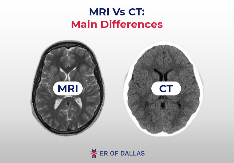
The core mechanism involves an X-ray beam passing through the body. Different tissues absorb X-rays to varying degrees: dense structures like bone absorb more, while softer tissues like organs absorb less. Detectors on the opposite side of the X-ray tube measure the amount of radiation that passes through. A powerful computer then processes these multiple “slices” of data, digitally reconstructing them into detailed cross-sectional images. Think of it as slicing a loaf of bread and then using a computer to create a 3D model of the whole loaf from those slices. The resulting images are displayed as gray-scale pictures, where brighter areas represent denser tissues (like bone) and darker areas represent less dense tissues (like air in the lungs). The real technological marvel here is the computational power required to synthesize these vast amounts of data into a coherent, multi-dimensional view of the body’s interior. Modern CT scanners boast incredible speed and resolution, thanks to continuous advancements in detector technology and processing algorithms.
Magnetic Resonance Imaging (MRI): Magnets, Radio Waves, and Hydrogen Atoms
MRI technology, in stark contrast, completely eschews ionizing radiation. Instead, it relies on a potent combination of a powerful magnetic field and radio waves to generate remarkably detailed images of organs, soft tissues, bone, and virtually all other internal body structures.
How it works:
The human body is largely composed of water molecules, and water molecules contain hydrogen atoms. Each hydrogen atom has a single proton, which acts like a tiny magnet, spinning and orienting itself randomly. The MRI scanner’s primary component is a super-strong magnet, typically thousands of times more powerful than a refrigerator magnet. When a patient enters the scanner, this magnetic field forces all the hydrogen protons in their body to align in the same direction, much like compass needles aligning with the Earth’s magnetic field.
Next, a brief pulse of radio waves is sent through the patient. This energy temporarily knocks the aligned hydrogen protons out of alignment. When the radiofrequency pulse is turned off, the protons “relax” back into alignment with the main magnetic field. As they do, they release energy, emitting a faint radio signal. Different tissues relax at different rates and emit signals of varying intensities. These subtle differences are detected by receiver coils in the MRI machine. A sophisticated computer then processes these unique signals, translating them into highly detailed cross-sectional images. The genius of MRI lies in its ability to differentiate between various soft tissues based on their water content and molecular environment, providing unparalleled contrast and clarity for structures like the brain, spinal cord, ligaments, and cartilage.
Diagnostic Applications: When to Use Which
Given their distinct technological underpinnings, CT and MRI excel in visualizing different types of tissues and conditions. The choice between them is a critical clinical decision, often guided by the specific diagnostic question.
CT Scan: Speed, Bones, and Acute Conditions
The strengths of CT technology make it the go-to choice for scenarios demanding speed, the visualization of dense structures, and the assessment of acute trauma. Its ability to quickly capture images of bone, blood vessels, and internal bleeding makes it invaluable in emergency medicine.
Primary applications include:
- Emergency Situations: For rapidly diagnosing severe head injuries (identifying hemorrhage or fractures), acute stroke, internal organ damage from trauma (e.g., car accidents), and appendicitis. Its speed is paramount when time is critical.
- Bone and Joint Imaging: Superior for detecting bone fractures, complex joint injuries, and assessing bone tumors or infections. It provides excellent detail of bony architecture.
- Chest and Abdominal Scans: Widely used for diagnosing lung conditions (pneumonia, emphysema, cancer), kidney stones, and evaluating various abdominal organs for tumors or abscesses. CT angiography, using contrast dye, can visualize blood vessels throughout the body for blockages or aneurysms.
- Cancer Staging: Often used to determine the extent of cancer, identifying tumor size, location, and spread to other organs or lymph nodes.
- Image-Guided Procedures: CT’s real-time imaging capability makes it useful for guiding biopsies, draining abscesses, or precisely targeting radiation therapy.
MRI Scan: Soft Tissue Mastery and Detailed Insights
MRI’s unparalleled ability to differentiate between subtle variations in soft tissues positions it as the superior choice for examining complex structures where high contrast is crucial. It’s often preferred for chronic conditions, neurological issues, and detailed evaluations.
Primary applications include:
- Brain and Spinal Cord: The gold standard for detecting brain tumors, strokes (especially in early stages), multiple sclerosis (MS) plaques, aneurysms, infections, and spinal cord injuries or disc herniations. Its ability to see through bone provides clear views of neurological tissue.
- Joints and Ligaments: Exceptional for diagnosing injuries to ligaments, tendons, cartilage, and menisci in joints like the knee, shoulder, and wrist. Sports medicine heavily relies on MRI for detailed damage assessment.
- Abdominal and Pelvic Organs: Provides highly detailed images of organs like the liver, kidneys, pancreas, and reproductive organs, superior for identifying certain types of tumors, cysts, or inflammation.
- Breast Imaging: In conjunction with mammography, MRI is used for breast cancer screening in high-risk women and for further evaluating abnormalities found on mammograms.
- Vascular Studies (MR Angiography): Can visualize blood vessels without the use of ionizing radiation, useful for assessing blockages or aneurysms.
Advantages and Disadvantages: Weighing the Choices
Both CT and MRI scans represent monumental achievements in medical imaging. However, each technology comes with its own set of trade-offs, making the selection process a careful balance of diagnostic need, patient safety, and practical considerations.
The CT Scan Perspective
Advantages:
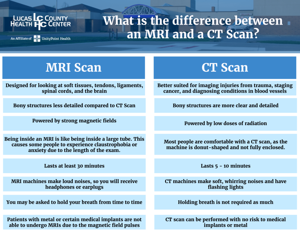
- Speed: CT scans are incredibly fast, often taking only a few minutes. This is critical in emergency situations where rapid diagnosis can be life-saving.
- Bone Detail: Offers superior resolution for bone structures, making it excellent for fractures, bone tumors, and intricate bony anatomy.
- Availability & Cost: CT scanners are more widely available and generally less expensive than MRI machines.
- Claustrophobia Friendly: The open design of many CT scanners is more comfortable for claustrophobic patients compared to the confined space of an MRI.
- Metal Implants: Generally safe for patients with certain metal implants that might be problematic for an MRI (e.g., pacemakers, some surgical clips, cochlear implants, though this always requires careful screening).
Disadvantages:
- Ionizing Radiation: The primary drawback is exposure to ionizing radiation, which carries a small, cumulative risk of cancer. While doses are carefully managed, this is a significant consideration, especially for pediatric patients or repeated scans.
- Limited Soft Tissue Contrast: While good, it doesn’t offer the same level of soft tissue differentiation as MRI, meaning subtle changes in organs or ligaments might be missed.
- Contrast Agent Risks: Iodine-based contrast dyes, sometimes used to enhance visibility of blood vessels or organs, can cause allergic reactions or kidney problems in susceptible individuals.
The MRI Scan Perspective
Advantages:
- No Ionizing Radiation: This is its most significant advantage, making it a safer option for patients who may require multiple scans, pregnant women (after careful consideration), and children.
- Exceptional Soft Tissue Contrast: Unrivaled in its ability to differentiate between various soft tissues, providing exquisite detail for conditions affecting the brain, spinal cord, muscles, ligaments, and internal organs.
- Versatility: Can provide different types of images by altering magnetic field gradients and radio pulse sequences, allowing radiologists to visualize different tissue properties (e.g., fluid-filled areas, fat, blood flow).
Disadvantages:
- Time-Consuming: MRI scans are significantly slower than CT scans, often taking 30 minutes to over an hour, which can be challenging for patients, especially those in pain or unable to lie still.
- Cost: Generally more expensive than CT scans due to the complex technology and infrastructure required.
- Claustrophobia: The enclosed, narrow tube of the MRI scanner can induce severe anxiety and claustrophobia in many patients. Open MRI machines exist but often have lower field strengths and therefore reduced image quality.
- Loud Noise: The rapid switching of gradient coils creates loud banging and knocking noises, necessitating ear protection.
- Magnetic Field Risks: The powerful magnet poses significant safety risks. Patients with certain metallic implants (e.g., older pacemakers, specific aneurysm clips, shrapnel) cannot undergo an MRI. Even non-ferromagnetic items must be removed from the scan room.
- Motion Sensitivity: Image quality can be significantly degraded by patient movement.
Technological Evolution and Future Trends
The journey of medical imaging is far from over. Both CT and MRI technologies are continually evolving, pushed forward by advancements in computing, physics, and materials science, promising even more precise, safer, and patient-friendly diagnostics.
Enhancements in CT Technology
Modern CT scanners are vastly superior to their predecessors. Innovations are primarily focused on reducing radiation dose while improving image quality and speed:
- Lower-Dose Protocols: Techniques like iterative reconstruction and dose modulation automatically adjust radiation output, significantly lowering patient exposure without compromising diagnostic quality.
- Faster Scanners: Multi-slice CT (MSCT) and spiral CT allow for quicker data acquisition, meaning full body scans can be done in seconds, reducing motion artifacts and improving throughput.
- AI-Powered Reconstruction: Artificial intelligence is increasingly used to reconstruct images from less data, further reducing radiation dose and enhancing image sharpness.
- Photon-Counting CT: An emerging technology that offers unprecedented resolution and material differentiation by individually counting X-ray photons, promising a new era of ultra-detailed CT imaging.
Advancements in MRI Technology
MRI research is exploring higher field strengths, enhanced patient comfort, and new functional capabilities:
- Higher Field Strengths: Clinical 3 Tesla (3T) MRI scanners are becoming more common, offering superior signal-to-noise ratio and thus higher resolution than standard 1.5T machines. Research 7T scanners provide even more exquisite detail, pushing the boundaries of neurological and musculoskeletal imaging.
- Silent MRI: Technologies are being developed to significantly reduce the characteristic loud noises of an MRI, making the experience more tolerable for patients.
- Faster Sequences: New pulse sequences and parallel imaging techniques are reducing scan times, addressing one of MRI’s key drawbacks.
- Functional MRI (fMRI): This specialized MRI measures brain activity by detecting changes in blood flow, providing insights into neurological function, research into cognitive processes, and surgical planning.
- AI for Image Analysis and Acquisition: AI algorithms are being deployed to optimize image acquisition parameters, correct for motion, and assist radiologists in interpreting complex scans, improving diagnostic accuracy and efficiency.
Complementary Roles: Integration for Comprehensive Diagnostics
Perhaps the most significant trend is not in pitting CT against MRI, but in understanding their complementary roles. Often, a patient might undergo both a CT and an MRI scan to get the most comprehensive picture. For example, a patient with a head injury might first receive a rapid CT scan to rule out acute bleeding or fracture, followed by an MRI if soft tissue damage or subtle neurological issues are suspected. AI tools are also emerging that can help clinicians navigate the optimal imaging pathway, suggesting the most appropriate scan based on symptoms, patient history, and initial findings, ensuring the most effective and safest diagnostic approach.
Patient Experience and Safety Considerations
While the technological prowess of CT and MRI is astounding, the patient’s experience and safety remain paramount. Understanding what to expect and the inherent risks is crucial for informed decision-making.
Preparing for Your Scan
Preparation varies depending on the type of scan and whether contrast agents are used. Generally, patients may be asked to fast for a few hours prior to a scan requiring contrast. Loose, comfortable clothing is recommended, and all metallic objects (jewelry, watches, glasses, dentures, hearing aids) must be removed, especially for an MRI due to its powerful magnetic field. For MRI, a thorough screening for internal metal implants is mandatory. Patients who are claustrophobic might be offered a mild sedative for an MRI.
Understanding the Risks
- CT: The primary concern is the ionizing radiation. While the risk of developing cancer from a single scan is very small, cumulative exposure from multiple scans over a lifetime is a consideration. Radiologists and technologists meticulously follow ALARA (As Low As Reasonably Achievable) principles to minimize dose. Contrast reactions, though rare, are also a risk.
- MRI: The main risks stem from the powerful magnetic field interacting with metal objects. Unscreened metallic implants can heat up, move, or malfunction, potentially causing serious injury. Patients with pacemakers or certain cerebral aneurysm clips are typically excluded. Acoustic noise can cause temporary hearing loss without protection. The contained environment can also be a source of psychological distress for some.
In conclusion, CT and MRI scans stand as monumental achievements in medical technology, each leveraging distinct physical principles to offer invaluable insights into the human body. The CT, with its reliance on X-rays, excels in speed and bone detail, making it indispensable for acute trauma and emergency diagnostics. The MRI, harnessing magnetic fields and radio waves, shines with unparalleled soft tissue contrast, critical for neurological, musculoskeletal, and organ-specific evaluations. As technology continues to advance, these powerful tools will only become more refined, safer, and more integrated, collectively enhancing our ability to diagnose, monitor, and treat an ever-expanding array of medical conditions, firmly cementing their place as cornerstones of modern medical tech.
aViewFromTheCave is a participant in the Amazon Services LLC Associates Program, an affiliate advertising program designed to provide a means for sites to earn advertising fees by advertising and linking to Amazon.com. Amazon, the Amazon logo, AmazonSupply, and the AmazonSupply logo are trademarks of Amazon.com, Inc. or its affiliates. As an Amazon Associate we earn affiliate commissions from qualifying purchases.