The question of what constitutes a “normal” uterine size is fundamentally a biological and medical one, central to women’s reproductive health. However, in the 21st century, the ability to accurately answer this question, to monitor deviations, and to provide comprehensive care is inextricably linked to technological advancement. From sophisticated imaging devices to artificial intelligence and remote monitoring solutions, technology has transformed our understanding and management of uterine health. This article delves into the technological landscape that enables medical professionals and individuals to gain insights into uterine dimensions, making it possible to define, detect, and address what is normal and when intervention might be necessary. It’s through these digital lenses and smart algorithms that we gain a precise picture of this vital organ, ensuring better diagnostic accuracy, personalized care, and empowered patient engagement.
The Digital Lens: Imaging Technologies Revolutionizing Uterine Assessment
At the forefront of understanding uterine size and morphology are advanced medical imaging technologies. These tools provide non-invasive, detailed visualizations of the uterus, allowing clinicians to measure its dimensions, assess its structure, and identify any anomalies. The evolution of these technologies has moved from rudimentary observational methods to highly precise digital reconstructions, dramatically improving diagnostic capabilities.
Ultrasound: The First Glimpse
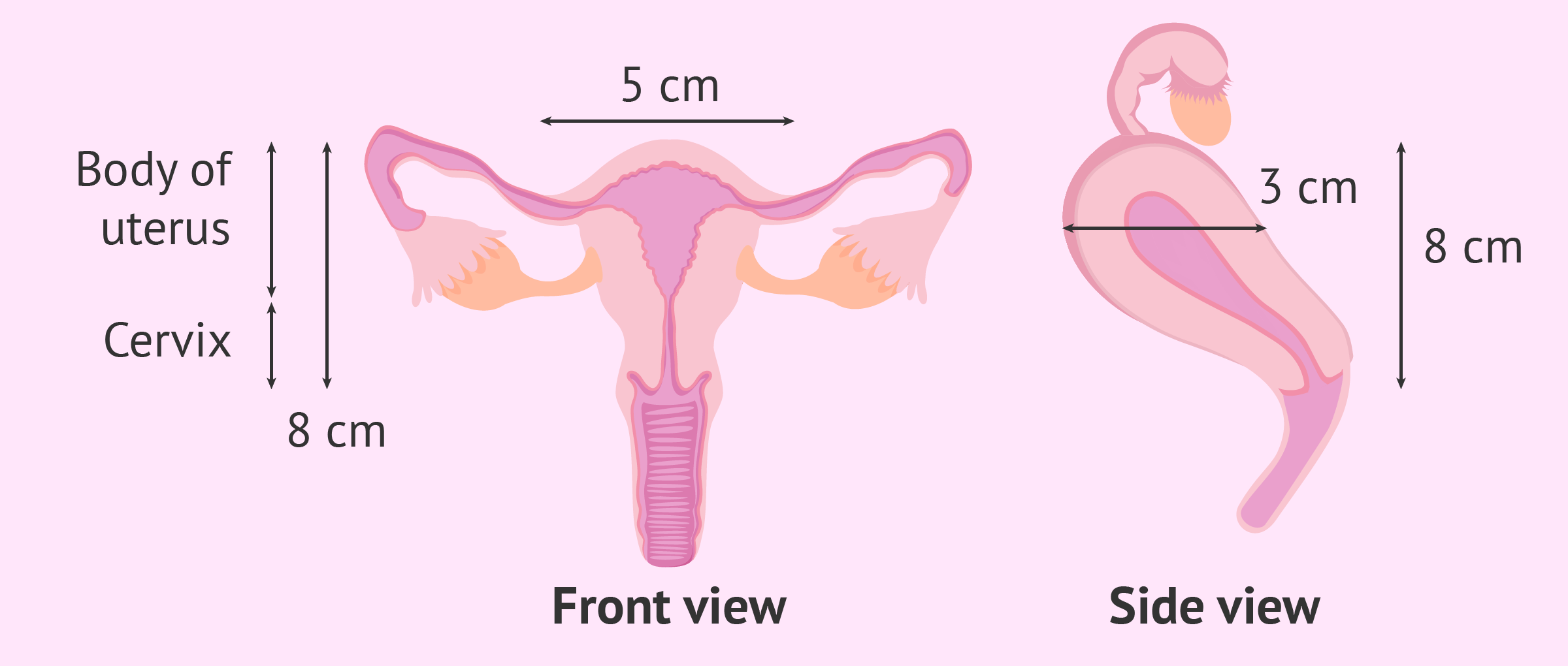
Ultrasound technology remains the cornerstone for assessing uterine size and structure. Utilizing high-frequency sound waves, an ultrasound transducer generates real-time images of internal organs. For uterine assessment, both transabdominal and transvaginal approaches are commonly employed. Transvaginal ultrasound, in particular, offers superior resolution due to its proximity to the uterus, providing clear measurements of uterine length, width, and anteroposterior diameter.
The digital advancements in ultrasound machines have been profound. Modern systems feature high-definition transducers, sophisticated signal processing, and intuitive user interfaces. This enables clinicians to capture highly detailed images, perform precise caliper measurements, and store data digitally for longitudinal tracking. Beyond basic measurements, advanced Doppler capabilities can assess blood flow to and within the uterus, offering additional insights into conditions like adenomyosis or fibroids. The immediate, non-invasive, and relatively inexpensive nature of ultrasound makes it the first-line diagnostic tool for assessing uterine size and detecting common abnormalities.
MRI and CT: Precision and Detail
While ultrasound provides excellent initial insights, Magnetic Resonance Imaging (MRI) and Computed Tomography (CT) scans offer even greater detail and are often used for more complex cases or when ultrasound findings are ambiguous.
MRI uses a powerful magnetic field and radio waves to create detailed images of organs and soft tissues. It excels at differentiating between various soft tissue types, making it invaluable for precisely measuring uterine size, identifying the exact location and size of fibroids (leiomyomas), adenomyosis, or congenital uterine anomalies. MRI provides multi-planar views without ionizing radiation, offering superior tissue contrast compared to ultrasound. Digital MRI platforms now integrate advanced sequencing and post-processing tools, allowing for 3D reconstructions and quantitative analysis of uterine volume and lesion characteristics.
CT scans, on the other hand, use X-rays to create cross-sectional images. While less commonly used specifically for uterine size assessment due to radiation exposure and less soft-tissue differentiation than MRI, CT can provide valuable information when assessing pelvic masses or in emergency situations where a rapid, comprehensive view of the abdominal and pelvic organs is needed. Digital CT scanners with multi-detector arrays offer faster acquisition times and thinner slices, enabling detailed reconstructions that contribute to a holistic understanding of the pelvic anatomy, including uterine position relative to other structures.
3D and 4D Imaging: Enhanced Visualization
Building upon conventional 2D imaging, 3D and 4D ultrasound technologies have introduced a new dimension of visualization for uterine assessment. 3D ultrasound acquires multiple 2D images and reconstructs them into a three-dimensional volume, allowing clinicians to view the uterus from any angle. This is particularly useful for evaluating uterine shape, identifying septa or bicornuate uteri, and precisely mapping the spatial relationship of fibroids or polyps. The ability to manipulate and rotate the 3D rendering on a digital workstation provides unparalleled insights into the uterine cavity and outer contour.
4D ultrasound adds the element of time to 3D imaging, essentially providing real-time 3D images. While more commonly associated with fetal imaging, 4D capabilities can be beneficial for visualizing dynamic processes within the uterus, though its primary utility for routine uterine size assessment is limited compared to its diagnostic power for structural anomalies. The software and processing power behind these advanced imaging modalities are crucial, enabling rapid acquisition, sophisticated reconstruction algorithms, and seamless integration into digital patient records.
AI and Machine Learning: Automating Uterine Diagnostics and Data Analysis
The sheer volume of data generated by advanced imaging techniques presents both an opportunity and a challenge. This is where Artificial Intelligence (AI) and Machine Learning (ML) step in, transforming how medical images are interpreted, diagnoses are made, and patient outcomes are predicted. AI tools are becoming indispensable in enhancing accuracy, efficiency, and personalization in uterine health assessment.
AI in Image Interpretation
AI-powered algorithms are being trained on vast datasets of ultrasound, MRI, and CT images of the uterus. These algorithms can identify and segment the uterus, measure its dimensions (length, width, depth, volume) with remarkable precision, and detect abnormalities such as fibroids, polyps, or adenomyosis. For instance, an AI system can be trained to automatically identify and quantify fibroids, even small ones, distinguishing them from surrounding healthy tissue. This capability significantly reduces the time radiologists and gynecologists spend on manual measurements and interpretation, minimizes inter-observer variability, and potentially catches subtle findings that might be overlooked by the human eye.
Furthermore, AI can assist in the classification of uterine anomalies, comparing a patient’s uterine morphology against a database of “normal” and “abnormal” patterns, helping clinicians arrive at a more accurate diagnosis faster. This augmentation of human expertise with AI’s analytical power marks a significant leap in diagnostic precision.

Predictive Analytics for Uterine Health
Beyond diagnosis, AI and ML are paving the way for predictive analytics in uterine health. By analyzing a multitude of factors—including imaging data, patient history, genetic markers, and lifestyle information—AI models can predict the likelihood of developing certain uterine conditions, the growth rate of fibroids, or the potential response to different treatments. For example, machine learning algorithms could predict which patients with uterine fibroids are most likely to benefit from a particular surgical procedure versus non-surgical management, optimizing treatment pathways.
These predictive models are continuously refined through exposure to new data, leading to increasingly accurate forecasts. This proactive approach allows for earlier intervention, personalized risk assessment, and more informed patient counseling, moving from reactive treatment to preventive and personalized care strategies.
Personalized Medicine and AI-Driven Insights
The ultimate goal of AI in uterine health is to facilitate personalized medicine. By integrating and analyzing diverse data points—from genomic information to detailed imaging and lifestyle data—AI can create a holistic profile for each patient. This enables clinicians to understand how a patient’s unique biological makeup and external factors influence their uterine health.
AI-driven insights can help determine a patient’s individual “normal” range for uterine size, factoring in age, parity, hormonal status, and ethnic background, which might deviate from population averages. This personalized approach moves away from a one-size-fits-all definition of normal, allowing for more nuanced diagnoses and highly tailored treatment plans, ultimately leading to better health outcomes and a more precise understanding of an individual’s reproductive health trajectory.
Wearables and Remote Monitoring: Empowering Proactive Uterine Health Management
The revolution in health technology extends beyond the clinic, enabling individuals to actively participate in monitoring their own health. Wearable devices, digital health apps, and telemedicine platforms are empowering proactive management of reproductive health, offering new ways to track cycles, identify symptoms, and connect with healthcare providers remotely. While not directly measuring uterine size, these technologies provide crucial contextual data and facilitate early detection of issues that might impact uterine health.
Smart Devices for Reproductive Health Tracking
An increasing array of smart devices and mobile applications are designed to track menstrual cycles, ovulation, and other reproductive health indicators. While devices like smart rings or watches might monitor basal body temperature, heart rate, and sleep patterns, dedicated apps allow users to log symptoms, discharge, and cycle length. These data points, when collected over time, can reveal patterns and deviations that might signal underlying uterine issues. For example, consistently heavy or painful periods tracked through an app might prompt an individual to seek medical advice, potentially leading to early diagnosis of conditions like fibroids or endometriosis, which can affect uterine size and function.
The integration of these devices with health platforms allows for the aggregation of personal health data, providing a more comprehensive view of an individual’s reproductive health narrative. While not measuring the uterus directly, the trends identified by these tools can be a critical trigger for further investigation using clinical imaging technologies.
Telemedicine and Virtual Consultations
Telemedicine platforms have rapidly evolved, particularly in recent years, to provide virtual healthcare consultations. For women’s reproductive health, this means individuals can discuss symptoms, review imaging results, and receive preliminary advice from gynecologists or other specialists without needing an in-person visit. While a remote consultation cannot replace a physical examination or imaging, it can facilitate initial screenings, follow-up appointments, and management of chronic conditions.
For questions regarding uterine size or related concerns, telemedicine can enable a specialist to review digitally transmitted ultrasound or MRI images, provide interpretations, and discuss findings with the patient. This enhances accessibility to expert care, reduces geographical barriers, and allows for more frequent and timely communication between patients and providers, ensuring that concerns related to uterine health are addressed promptly.
Data Security and Privacy in Women’s Health Tech
As technology increasingly integrates into sensitive areas of health, particularly reproductive health, the paramount importance of data security and patient privacy cannot be overstated. The digital transformation in uterine health diagnostics and management relies heavily on the secure handling of highly personal medical information, making robust cybersecurity measures and stringent privacy protocols non-negotiable.
Safeguarding Sensitive Health Information
The collection, storage, and transmission of medical images, AI-driven diagnostic reports, and personal health data from wearables raise significant privacy concerns. Healthcare technology providers must implement advanced encryption protocols, multi-factor authentication, and secure cloud storage solutions to protect this sensitive information from unauthorized access, breaches, or misuse. Data anonymization and pseudonymization techniques are crucial when using aggregated data for research or AI model training, ensuring individual patient identities are protected.
Patients need assurances that their reproductive health data, which can be particularly vulnerable to discrimination or other negative consequences if exposed, is handled with the utmost care and confidentiality. Trust in these systems is foundational to their adoption and effectiveness.

Regulatory Frameworks and Compliance
The landscape of health tech is governed by a complex web of regulatory frameworks, such as HIPAA in the United States, GDPR in Europe, and similar legislation worldwide. These regulations mandate strict standards for data privacy, security, and integrity. Technology companies developing solutions for uterine health must ensure full compliance with these laws, which typically cover informed consent for data collection, patient rights to access and correct their data, and strict accountability for data breaches.
Furthermore, as AI tools become more prevalent in diagnostics, regulatory bodies are also scrutinizing the ethical implications and algorithmic bias in these systems. Ensuring that AI models are trained on diverse datasets to prevent biased outcomes and that their decision-making processes are transparent and explainable is critical for responsible innovation in women’s health technology. Adherence to these frameworks not only ensures legal compliance but also builds essential patient and professional confidence in the digital future of uterine health.
In conclusion, while the question “what is the normal size for a uterus?” remains a medical inquiry, the ability to answer it, understand its nuances, and manage related conditions is increasingly mediated and enhanced by technology. From high-resolution imaging that precisely measures dimensions, to AI that interprets vast datasets and predicts outcomes, and wearables that empower personal health tracking, technology is redefining how we approach women’s reproductive health. This digital evolution is leading to more accurate diagnoses, personalized care, and a proactive approach to well-being, all while navigating the critical need for robust data security and privacy.
aViewFromTheCave is a participant in the Amazon Services LLC Associates Program, an affiliate advertising program designed to provide a means for sites to earn advertising fees by advertising and linking to Amazon.com. Amazon, the Amazon logo, AmazonSupply, and the AmazonSupply logo are trademarks of Amazon.com, Inc. or its affiliates. As an Amazon Associate we earn affiliate commissions from qualifying purchases.