In the vast and intricate world revealed by microscopy, few concepts are as fundamental and critically misunderstood as “resolving power.” Often conflated with mere magnification, a microscope’s resolving power—or resolution—is the true benchmark of its capability, determining its ability to distinguish between two closely spaced points as separate entities. It is the very essence of clarity, dictating how much detail we can discern in the minute structures of the universe, from the organelles within a cell to the atomic lattice of a material. For anyone venturing into scientific research, quality control, or advanced technological development, understanding this crucial technological specification is not just beneficial but indispensable. Without high resolving power, even the most powerful magnification would yield nothing more than a blurry, enlarged image, obscuring the very insights we seek to uncover.

The Fundamental Concept of Resolving Power
To truly appreciate the significance of resolving power, we must first disentangle it from its often-confused counterpart: magnification. While both are essential aspects of microscopy, their roles and implications are distinct.
Defining Resolution vs. Magnification
Magnification refers to the process of enlarging an image of something. If a microscope has a total magnification of 1000x, it means the image appears 1000 times larger than the actual object. This is intuitively understood and easily achieved; simply adding more lenses can increase magnification. However, there’s a critical limitation: beyond a certain point, increasing magnification without a corresponding increase in resolution simply results in a larger, but progressively blurrier, image. It’s like zooming in on a low-resolution digital photograph – you only magnify the pixels, not gain new detail.
Resolution, on the other hand, is the ability to distinguish two separate points or objects that are very close together. A microscope with high resolving power can show two adjacent bacteria as distinct entities, rather than a single, elongated blob. This ability is what truly reveals the intricate details of a specimen, allowing scientists to differentiate between subcellular structures, examine material defects, or identify individual viral particles. It is the gateway to new discoveries, providing the clarity necessary for meaningful observation and analysis.
The Abbe Diffraction Limit
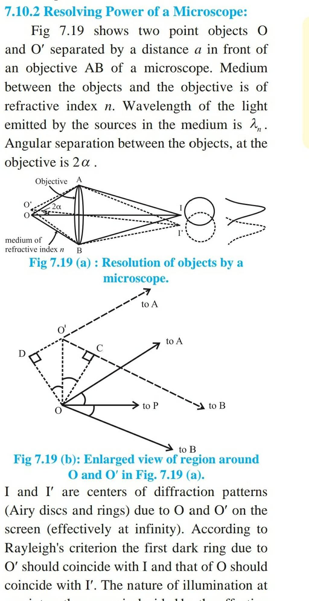
The theoretical maximum resolving power of a light microscope is governed by a fundamental principle of physics known as the Abbe Diffraction Limit, named after German physicist Ernst Abbe. This limit states that it is impossible to resolve details smaller than about half the wavelength of the light used for imaging.
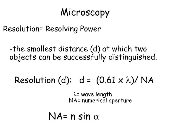
Abbe’s formula for the theoretical resolving power (d) of a microscope is:
d = (0.61 * λ) / NA
Where:
- d represents the minimum resolvable distance between two points (a smaller ‘d’ means better resolution).
- λ (lambda) is the wavelength of the light used to illuminate the specimen.
- NA is the numerical aperture of the objective lens.
This equation reveals the two primary factors that dictate the resolution capabilities of a traditional optical microscope: the wavelength of light and the numerical aperture of the objective lens. Understanding these components is key to maximizing a microscope’s performance and pushing the boundaries of what can be observed.
Factors Influencing a Microscope’s Resolving Power
Several critical factors contribute to or limit the resolving power of a microscope. Engineers and scientists meticulously optimize these elements to achieve the clearest possible images.
Wavelength of Light
As indicated by Abbe’s formula, the wavelength of the illuminating light (λ) is directly proportional to the minimum resolvable distance. This means that shorter wavelengths lead to better resolution. Visible light ranges from approximately 400 nm (violet) to 700 nm (red). Blue light, with its shorter wavelength (around 450-495 nm), provides better resolution than red light. This is why many high-resolution light microscopes utilize filters to favor shorter wavelengths or employ specialized light sources.
Pushing this concept further, electron microscopes achieve significantly higher resolution precisely because they use electrons instead of photons. Electrons, when accelerated to high velocities, exhibit wave-like properties with wavelengths thousands of times shorter than visible light, thereby dramatically reducing ‘d’ and enabling atomic-level resolution.
Numerical Aperture (NA) of the Objective Lens
The Numerical Aperture (NA) is arguably the most crucial factor dictated by the microscope’s optical design. It is a dimensionless number that quantifies the ability of an objective lens to gather light and resolve fine specimen detail. A higher NA indicates a greater light-gathering capability and, consequently, better resolution.
The formula for NA is:
NA = n * sin(µ)
Where:
- n is the refractive index of the medium between the objective lens and the specimen (e.g., air, water, oil).
- µ (mu) is the half-angle of the maximum cone of light that can enter the objective lens from the specimen (also known as the aperture angle).
A larger aperture angle (meaning a larger objective lens opening or a shorter working distance) allows more diffracted light to be collected, capturing more information and thus improving resolution.
Refractive Index of the Medium
The refractive index (n) of the medium between the specimen and the objective lens plays a direct role in the NA calculation. Air has a refractive index of approximately 1.0. However, using immersion oil (with a refractive index similar to glass, typically around 1.5) between the objective lens and the coverslip drastically increases the effective NA. This is because immersion oil prevents the refraction (bending) of light away from the objective lens that would otherwise occur at the air-glass interface, allowing more light rays to enter the lens. This is why high-magnification, high-resolution objective lenses are almost invariably designed for oil immersion.
Specimen Preparation and Contrast
While not directly part of the Abbe limit, the way a specimen is prepared significantly impacts the perceived resolution and the ability to distinguish structures. Poorly stained, thick, or unevenly mounted specimens can scatter light or obscure details, making it difficult for even a high-resolution microscope to provide a clear image. Techniques like staining, phase contrast, differential interference contrast (DIC), and fluorescence microscopy are employed to enhance the contrast of otherwise transparent biological samples, making their internal structures discernible.
Why Resolving Power Matters in Technology and Science
The relentless pursuit of higher resolving power has been a driving force behind countless scientific breakthroughs and technological advancements across diverse fields.
Advancements in Biological Research
In biology and medicine, improved resolution has revolutionized our understanding of life. From observing the intricate structures within a cell (organelles like mitochondria, endoplasmic reticulum) to visualizing the interactions between proteins and DNA, high-resolution microscopy has been indispensable. It has enabled the study of pathogens, the visualization of neural networks, and the detailed analysis of tissue pathology, contributing to drug discovery, disease diagnosis, and our fundamental comprehension of life processes. The ability to resolve individual bacteria, viruses, or even molecular complexes directly impacts our capacity to combat disease and understand cellular functions.
Materials Science and Nanotechnology
For materials scientists, resolving power is crucial for examining the microstructure of materials, identifying defects, and understanding their properties at the nanoscale. From analyzing the crystal lattice of metals and ceramics to characterizing the surface topography of semiconductors and polymers, high-resolution imaging is key. In the burgeoning field of nanotechnology, where scientists manipulate matter at the atomic and molecular level, microscopes with ultra-high resolving power (like electron microscopes and atomic force microscopes) are the primary tools for designing, synthesizing, and verifying nanostructures and nanomaterials.
Quality Control and Industrial Applications
Beyond research, resolving power is a cornerstone of modern industrial quality control. In the microelectronics industry, for instance, high-resolution microscopes are used to inspect silicon wafers for manufacturing defects, verify circuit patterns, and ensure the integrity of microchips. In precision manufacturing, they allow for the detailed examination of machined surfaces, welds, and coatings, ensuring product reliability and performance. Failure analysis often relies on high-resolution imaging to pinpoint the exact cause of component failure, guiding design improvements and preventing future issues.
Medical Diagnostics
In clinical settings, pathology labs heavily rely on microscopes to diagnose diseases. High resolution is essential for identifying cancerous cells, analyzing tissue biopsies, and detecting microbial infections. Accurate and timely diagnosis often hinges on the ability to discern subtle morphological changes at the cellular level, which directly correlates with the microscope’s resolving power.
Overcoming the Diffraction Limit: Advanced Microscopy Techniques
For centuries, the Abbe diffraction limit defined the boundaries of what could be seen with light. However, the 20th and 21st centuries have witnessed extraordinary technological innovations that have either bypassed this limit entirely or cleverly circumvented it.
Electron Microscopy (TEM, SEM)
The most dramatic leap in resolving power came with the invention of electron microscopes. Instead of using photons, these instruments employ a beam of electrons, which have significantly shorter wavelengths than visible light. This allows electron microscopes to achieve resolutions thousands of times greater than light microscopes, enabling scientists to visualize structures down to the atomic level.
- Transmission Electron Microscopy (TEM) shoots electrons through a thinly sectioned specimen, revealing internal ultrastructure.
- Scanning Electron Microscopy (SEM) scans a focused electron beam across the surface of a specimen, creating detailed three-dimensional topographical images.
Electron microscopy revolutionized fields like material science, biology, and chemistry by unveiling previously invisible worlds.
Super-Resolution Light Microscopy (STED, PALM/STORM)
A more recent and groundbreaking development has been the advent of super-resolution light microscopy techniques. These methods cleverly circumvent the Abbe diffraction limit using light, earning their developers the Nobel Prize in Chemistry in 2014. Rather than relying on shorter wavelengths, these techniques manipulate the fluorescence of molecules to effectively localize their positions with much greater precision than the diffraction limit would normally allow.
- Stimulated Emission Depletion (STED) microscopy uses two laser beams: one to excite fluorescent molecules and another (a “depletion” beam) to switch off fluorescence in the periphery of the excited spot, effectively shrinking the fluorescent area and improving resolution.
- Photoactivated Localization Microscopy (PALM) / Stochastic Optical Reconstruction Microscopy (STORM) rely on the stochastic activation and precise localization of individual fluorescent molecules over time. By taking many images and computationally reconstructing the positions of millions of molecules, a high-resolution image is built up.
These techniques allow researchers to observe living cells and their dynamic processes at nanoscale resolution, bridging the gap between traditional light microscopy and electron microscopy.
Atomic Force Microscopy (AFM)
Atomic Force Microscopy (AFM) operates on a completely different principle, not using light or electrons. Instead, it uses a sharp probe mounted on a cantilever to “feel” the surface of a specimen. As the probe scans, van der Waals forces between the probe tip and the sample surface cause the cantilever to deflect. These deflections are measured and converted into a topographical map of the surface, achieving atomic-level resolution in the z-axis and sub-nanometer resolution laterally. AFM is particularly useful for studying surface characteristics of materials, biological samples, and even individual molecules in ambient conditions.
Future Trends and Innovations in Microscopy
The journey to higher resolving power and more insightful imaging is far from over. The field of microscopy continues to evolve rapidly, driven by interdisciplinary advancements in optics, materials science, and computational power.
AI-Powered Image Processing
Artificial intelligence and machine learning are increasingly being integrated into microscopy workflows. AI algorithms can be trained to de-noise images, enhance contrast, segment features, and even computationally reconstruct higher-resolution images from lower-resolution data. This not only improves the quality of existing microscopy techniques but also makes image analysis faster and more objective, pushing the boundaries of what can be extracted from raw microscopy data.
Miniaturization and Portability
The trend towards miniaturization is bringing high-performance microscopy out of specialized labs and into the field or even point-of-care settings. Compact, portable microscopes, some even smartphone-attachable, are being developed with surprisingly good resolving power. These innovations promise to democratize access to microscopic analysis, enabling rapid diagnostics, environmental monitoring, and educational outreach in remote areas.
Multi-Modal Imaging
Future microscopy will increasingly involve multi-modal approaches, combining different imaging techniques to gather complementary information from a single specimen. For example, integrating optical microscopy with electron microscopy or AFM, alongside spectroscopic techniques, can provide a holistic view of both the morphology and chemical composition of a sample at various scales. This comprehensive data acquisition will unlock deeper insights into complex biological systems and advanced materials.
Conclusion
The resolving power of a microscope is far more than a technical specification; it is the gateway to unseen worlds, a critical determinant of scientific discovery and technological innovation. From Abbe’s fundamental limit to the revolutionary leaps of electron microscopy and the ingenious circumventing strategies of super-resolution light microscopy, the continuous pursuit of greater clarity has reshaped our understanding of biology, materials, and the very fabric of existence. As technology continues to advance, fueled by AI, miniaturization, and multi-modal approaches, the future of microscopy promises even more breathtaking revelations, pushing the boundaries of human knowledge and enabling unprecedented insights into the infinitesimal details that govern our macroscopic world. For anyone engaged with technology, understanding and leveraging this power remains paramount.
aViewFromTheCave is a participant in the Amazon Services LLC Associates Program, an affiliate advertising program designed to provide a means for sites to earn advertising fees by advertising and linking to Amazon.com. Amazon, the Amazon logo, AmazonSupply, and the AmazonSupply logo are trademarks of Amazon.com, Inc. or its affiliates. As an Amazon Associate we earn affiliate commissions from qualifying purchases.