
In the intricate landscape of human physiology, the stomach plays a pivotal role in initiating digestion, acting as a temporary reservoir and processing unit for food before it moves into the small intestine. When this crucial process falters, leading to symptoms like nausea, vomiting, early satiety, or bloating, medical professionals often turn to advanced diagnostic tools to pinpoint the problem. Among these, the stomach emptying test, also known as a gastric emptying scintigraphy (GES), stands out as a highly specialized procedure. Far from a simple observation, this test is a testament to the remarkable advancements in medical technology, integrating principles of nuclear medicine, advanced imaging, and sophisticated data analysis to provide crucial insights into gastrointestinal motility.

This article will navigate the technological underpinnings of the stomach emptying test, exploring the sophisticated equipment, software, and methodologies that enable its precision. We will delve into how radiopharmaceutical tracers interact with gamma cameras, the role of digital processing in translating raw data into diagnostic insights, and the cutting-edge innovations that are continually refining this essential diagnostic tool, firmly positioning it within the realm of medical technology.
The Core Mechanism: Radiopharmaceutical Tracers and Imaging Technology
At its heart, the stomach emptying test is a triumph of engineering and radiochemistry, designed to non-invasively track the passage of food through the stomach. The procedure hinges on a carefully orchestrated interplay between a radioactive tracer, a specialized meal, and high-resolution imaging equipment.
How Radiotracers Work: A Glimpse into Nuclear Medicine
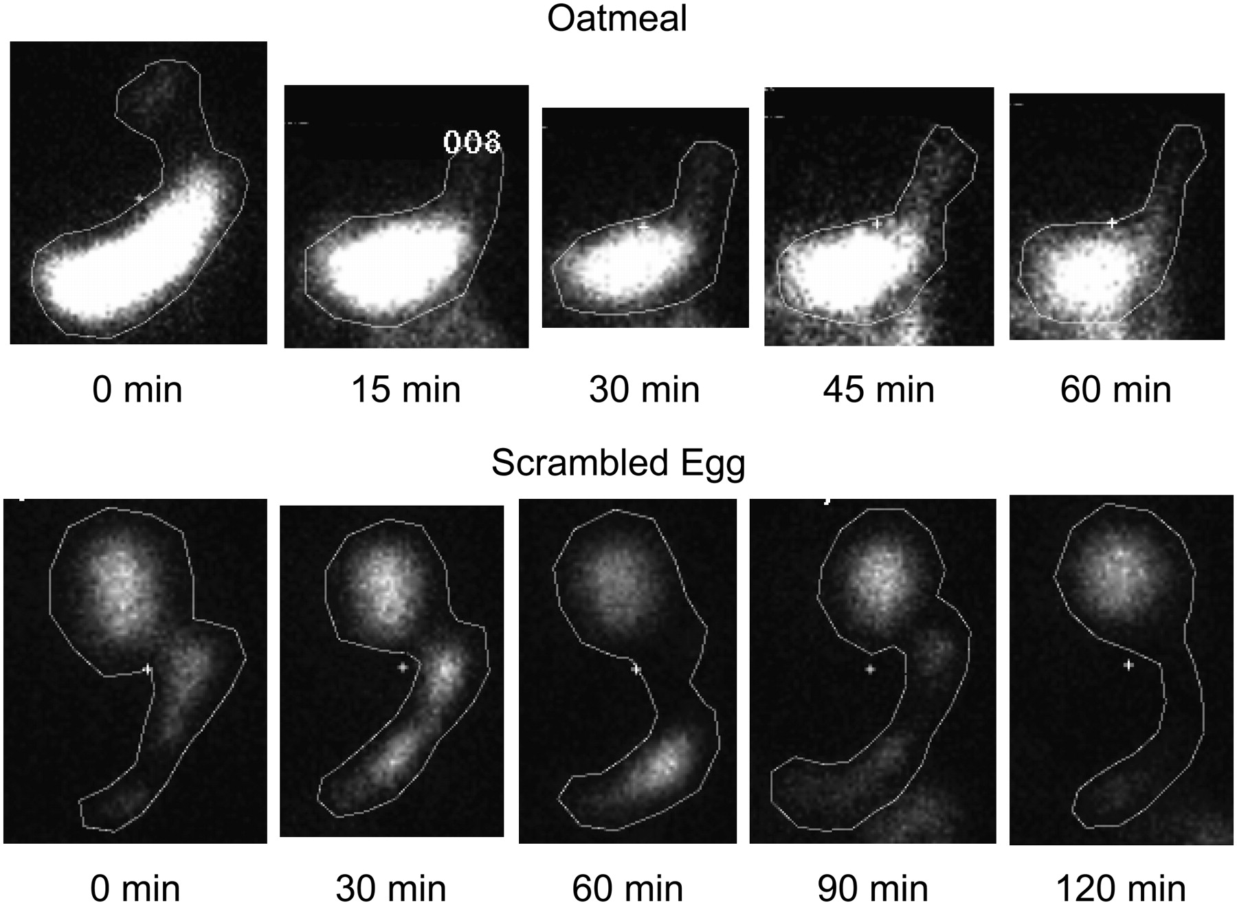
The test begins with the ingestion of a standardized meal, typically oatmeal or egg whites, that has been meticulously mixed with a small, harmless amount of a radiopharmaceutical—often Technetium-99m (Tc-99m). Tc-99m is chosen for its ideal properties: a short half-life (around 6 hours), ensuring minimal patient exposure to radiation, and its emission of gamma rays, which can be easily detected externally. Crucially, the radiotracer is bound to the solid component of the meal, as solid emptying rates are generally more indicative of gastric motility disorders than liquid emptying. Once ingested, the radiotracer emits gamma photons as it passes through the digestive tract, acting as a beacon for the imaging system. The precision in selecting and preparing this tracer is a fundamental technological consideration, balancing diagnostic efficacy with patient safety and minimizing artifact interference.
The Role of Gamma Cameras: Precision Imaging in Diagnostics
The detection of these gamma photons is the domain of the gamma camera, a cornerstone of nuclear medicine technology. A gamma camera, or Anger camera, consists of several key components working in concert. At its forefront is a collimator, a lead plate with thousands of tiny holes, which filters gamma rays, allowing only those traveling perpendicular to the detector to pass through. This ensures spatial resolution and image clarity. Behind the collimator lies a large thallium-activated sodium iodide crystal, which scintillates (emits light) when struck by a gamma photon. These light flashes are then detected by an array of photomultiplier tubes (PMTs), which convert the light signals into electrical pulses. The strength and location of these pulses are then processed to reconstruct a precise map of the radiotracer’s distribution within the stomach over time. The technological sophistication of gamma cameras, with their ability to capture subtle changes in radiotracer concentration, is paramount to the test’s diagnostic value. Modern gamma cameras are highly sensitive and capable of producing high-resolution images, essential for accurate gastric emptying curve generation.
Data Acquisition and Digital Processing: Translating Signals into Insights
The raw data collected by the gamma camera—thousands of electrical pulses corresponding to gamma photon detections—is then fed into a dedicated computer system. This is where digital signal processing truly shines. Specialized software algorithms are employed to process these signals, correct for background radiation, and compensate for patient movement, transforming ephemeral light flashes into quantifiable digital images. Multiple images are captured over several hours (typically 0, 1, 2, and 4 hours after meal ingestion), creating a time-series dataset. The software then defines regions of interest (ROIs) around the stomach and quantifies the remaining radioactivity within these ROIs at each time point. This sequential measurement allows for the generation of a gastric emptying curve, a graphical representation of the percentage of the meal remaining in the stomach over time. This curve is then compared against established normal ranges, allowing clinicians to diagnose conditions like gastroparesis (delayed emptying) or rapid gastric emptying. The accuracy of this digital processing, from image filtering to curve fitting, is critical for a reliable diagnosis.
Advancements in Stomach Emptying Test Technology
The foundational technology of the stomach emptying test, while robust, has not remained static. Continuous innovation in hardware and software has pushed the boundaries of diagnostic accuracy, efficiency, and patient experience.
From Planar to SPECT/CT: Enhancing Accuracy and 3D Visualization
Historically, gastric emptying studies primarily utilized planar imaging, providing a two-dimensional projection of the stomach’s contents. While effective, planar imaging can suffer from overlap of structures and less precise quantification due to attenuation of gamma rays by surrounding tissues. The advent of Single-Photon Emission Computed Tomography (SPECT) and its integration with Computed Tomography (CT) has revolutionized the field. SPECT/CT systems combine the functional information of SPECT (showing radiotracer distribution in 3D) with the anatomical precision of CT (showing detailed tissue structure). This fusion allows for highly accurate volumetric quantification of gastric contents and precise localization of the tracer, correcting for potential errors caused by overlapping organs or patient body habitus. The technological leap from 2D to 3D imaging, facilitated by sophisticated reconstruction algorithms and powerful computing, offers clinicians a more nuanced and reliable picture of gastric motility.
Artificial Intelligence and Machine Learning in Image Analysis
The sheer volume of data generated by advanced imaging techniques presents both a challenge and an opportunity. Artificial intelligence (AI) and machine learning (ML) are rapidly being integrated into the analysis of gastric emptying studies. AI algorithms can be trained on vast datasets of patient scans to automate the identification of regions of interest, segment organs, and even detect subtle anomalies that might be missed by the human eye. Machine learning models can analyze gastric emptying curves with greater precision, identifying patterns indicative of specific disorders and potentially predicting treatment responses. For instance, AI could assist in standardizing measurements across different institutions, reducing inter-observer variability, and speeding up the diagnostic process. This technological integration promises to enhance diagnostic accuracy, reduce workload for radiologists and nuclear medicine physicians, and unlock deeper insights from existing data.
Miniaturization and Portability: The Future of Diagnostic Devices

The trend towards miniaturization and portability, prevalent across many tech sectors, is also impacting medical diagnostics. While full gamma cameras remain large, research is exploring smaller, more portable detection systems, potentially leading to more flexible testing environments or even wearable solutions for continuous monitoring. Imagine a future where a patient could undergo a gastric emptying assessment with a compact device, transmitting data wirelessly to a cloud-based analytics platform. This would dramatically improve convenience for patients and reduce the logistical burden on healthcare facilities, making advanced diagnostics more accessible. Such innovations hinge on breakthroughs in detector materials, wireless communication protocols, and energy-efficient processing chips.
Software and Data Analytics: Beyond the Image
Beyond the physical hardware, the stomach emptying test is equally reliant on sophisticated software and robust data analytics frameworks that transform raw signals into actionable medical intelligence.
Specialized Software for Gastric Emptying Quantification
Dedicated software packages are the brains behind the operation, providing the tools for image reconstruction, quantification, and curve generation. These programs are meticulously designed to handle the unique requirements of nuclear medicine imaging. They offer features such as automatic ROI definition, attenuation correction, motion correction, and various mathematical models for curve fitting (e.g., linear, exponential, power exponential). The user interfaces are designed to be intuitive for medical professionals, allowing for efficient workflow and robust quality control. The development of such specialized software involves complex algorithmic design, adherence to strict medical device regulations, and continuous updates to incorporate new research findings and technological improvements.
Predictive Analytics and Personalized Medicine
The data generated from gastric emptying tests, especially when combined with other patient data (demographics, symptoms, co-morbidities, genetic markers), holds immense potential for predictive analytics. Machine learning models can analyze these multi-modal datasets to predict which patients are more likely to respond to certain treatments, identify subgroups with specific disease phenotypes, or even forecast disease progression. This moves diagnostics beyond mere identification of a problem towards a more proactive, personalized medicine approach. For example, understanding a patient’s unique gastric emptying pattern through advanced analytics could guide drug selection and dosage, optimizing therapeutic outcomes and minimizing adverse effects, a true technological leap in patient care.
Integration with Electronic Health Records (EHRs) for Seamless Data Flow
The digital nature of stomach emptying test results makes them ideal candidates for integration with Electronic Health Record (EHR) systems. Seamless integration ensures that the detailed gastric emptying curves, images, and interpretation reports are immediately accessible to all members of the patient’s care team. This eliminates delays, reduces the risk of transcription errors, and facilitates a holistic view of the patient’s health. Technologies like Health Level Seven International (HL7) and Fast Healthcare Interoperability Resources (FHIR) standards are critical for enabling this interoperability, allowing diverse medical systems to communicate effectively. This interconnectedness is a cornerstone of modern digital healthcare, enhancing decision-making and improving coordination of care.
Emerging Technologies and Future Directions
The pace of technological innovation ensures that the field of gastric motility diagnostics will continue to evolve, with several exciting avenues currently under exploration.
Smart Pills and Wireless Monitoring Systems
One of the most promising areas is the development of ingestible “smart pills.” These miniature electronic devices, equipped with sensors, cameras, and even wireless transmitters, can measure various physiological parameters, including pH, temperature, and pressure, as they traverse the gastrointestinal tract. While not directly replacing gastric emptying scintigraphy yet, certain smart pill technologies are being developed to measure transit times, offering a non-radioactive alternative for specific applications. Continuous, wireless monitoring via ingestible sensors could provide a more dynamic and prolonged assessment of gastric motility than a single-time-point scintigraphy, potentially revealing patterns that current tests might miss. This represents a significant shift towards less invasive and more patient-friendly diagnostic tools, driven by microelectronics and wireless communication tech.
Non-Radioactive Alternatives: Breath Tests and MRI Applications
The inherent use of radiation, however minimal, in scintigraphy prompts continuous research into non-radioactive alternatives. Breath tests, for instance, utilize a non-radioactive isotope of carbon (C-13) incorporated into a meal. As the meal is digested and emptied, the C-13 is metabolized and released in the breath as C-13 carbon dioxide, which can be measured. Magnetic Resonance Imaging (MRI) is another powerful non-ionizing imaging modality being explored for gastric emptying assessment. Functional MRI techniques can visualize and quantify gastric volume changes over time with high spatial resolution, offering a comprehensive view without radiation exposure. These alternatives, while still evolving, demonstrate the ongoing technological quest for equally accurate but safer diagnostic methods.
Cybersecurity in Medical Diagnostics: Protecting Sensitive Patient Data
As medical diagnostics become increasingly digitized and interconnected, the importance of cybersecurity cannot be overstated. The data generated by stomach emptying tests, including patient identifiers, medical images, and diagnostic reports, are highly sensitive. Robust cybersecurity protocols, encryption standards, secure cloud storage, and stringent access controls are essential to protect this information from unauthorized access, breaches, and cyber threats. Ensuring the integrity and confidentiality of medical data is a critical technological challenge that underpins the trustworthiness and reliability of all modern digital diagnostic tools.
In conclusion, the stomach emptying test is far more than a simple medical procedure; it is a sophisticated application of diverse technologies, from nuclear physics and advanced imaging to complex software algorithms and artificial intelligence. Its evolution reflects the broader trajectory of medical diagnostics—a continuous push for greater precision, efficiency, safety, and integration. As technology continues to advance, we can anticipate even more innovative, patient-friendly, and insightful tools emerging to decode the mysteries of gastric motility, further solidifying the test’s place at the forefront of medical diagnostic technology.
aViewFromTheCave is a participant in the Amazon Services LLC Associates Program, an affiliate advertising program designed to provide a means for sites to earn advertising fees by advertising and linking to Amazon.com. Amazon, the Amazon logo, AmazonSupply, and the AmazonSupply logo are trademarks of Amazon.com, Inc. or its affiliates. As an Amazon Associate we earn affiliate commissions from qualifying purchases.