The femur, or thigh bone, is the longest and strongest bone in the human body, playing a crucial role in weight-bearing and mobility. A fracture in this vital bone, known as a femur bone fracture, is a serious injury that can significantly impact an individual’s life. While the immediate concern is often the pain and loss of function, understanding the mechanics, causes, symptoms, and treatment options for a femur fracture is essential for anyone seeking information on this complex medical issue. This article aims to demystify femur fractures, providing a clear and accessible overview for a general audience.
Understanding the Anatomy and Mechanics of the Femur
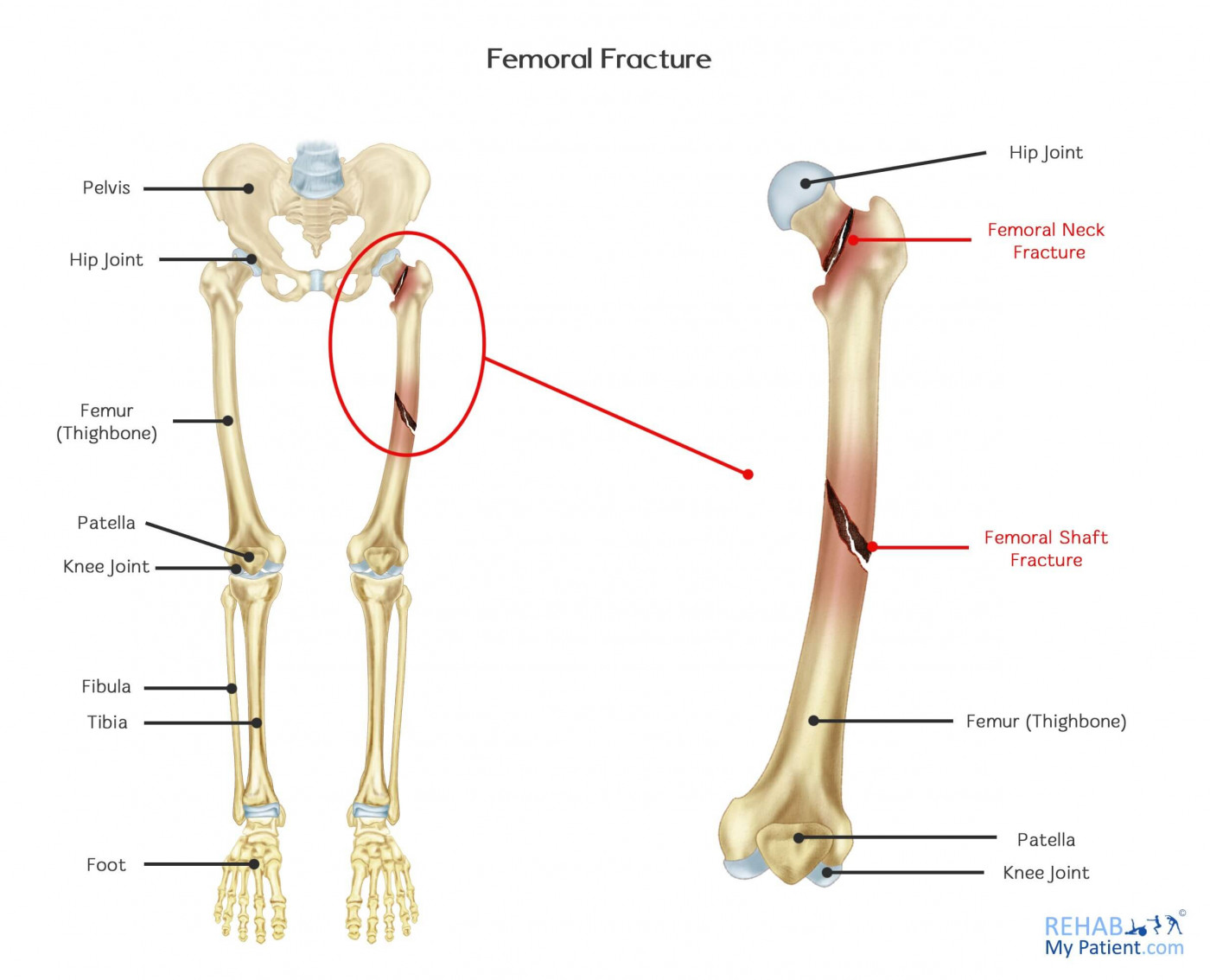
To grasp the implications of a femur fracture, it’s helpful to first understand the bone itself and its role in our skeletal system. The femur extends from the hip joint to the knee joint. Its proximal end connects to the pelvis at the hip socket (acetabulum), forming the hip joint. This articulation allows for a wide range of motion in the leg. The distal end of the femur articulates with the tibia (shinbone) and the patella (kneecap) at the knee joint, enabling us to bend and straighten our leg.
The femur is not a simple, solid rod. Its structure is remarkably engineered for strength and resilience. The shaft, or diaphysis, is a long, cylindrical portion, while the proximal and distal ends are expanded to form articular surfaces. The bone is also surrounded by robust muscles, most notably the quadriceps femoris group on the anterior (front) side and the hamstrings on the posterior (back) side. These muscles provide dynamic stability to the femur and are powerful force generators, essential for walking, running, and jumping.
The strength of the femur is further enhanced by its dense cortical bone, which forms a hard outer shell, and its porous, trabecular bone on the inside, which provides a lightweight yet strong internal structure. This intricate design allows the femur to withstand significant forces, including those generated by everyday activities and even moderate impacts. However, certain high-energy events or conditions that weaken the bone can lead to a fracture.
Causes and Mechanisms of Femur Fractures
Femur fractures are typically the result of significant trauma. The force required to break this robust bone is substantial, and understanding the mechanisms of injury can help in prevention and diagnosis. The most common causes of femur fractures can be broadly categorized:
High-Energy Trauma
This is by far the most prevalent cause, particularly in younger, healthier individuals. These incidents involve substantial forces applied to the femur, overwhelming its inherent strength.
- Motor Vehicle Accidents (MVAs): Collisions, especially those involving direct impact to the thigh or severe forces transmitted through the legs, are a leading cause. The speed and type of vehicle, as well as the nature of the impact, significantly influence the severity of the fracture. Dashboard injuries, where the knee strikes the dashboard during a frontal collision, are a common mechanism for distal femur fractures.
- Falls from Height: Significant falls, such as from a ladder, roof, or upper-story window, can generate forces sufficient to fracture the femur. The impact surface and the body’s landing position play a crucial role.
- Contact Sports and High-Impact Activities: While less common than MVAs, severe injuries in sports like football, rugby, or skiing, particularly those involving direct blows to the thigh or twisting forces, can lead to femur fractures.
Low-Energy Trauma and Underlying Bone Conditions
In older adults or individuals with compromised bone health, even relatively minor forces can lead to a femur fracture.
- Falls (especially in the Elderly): Osteoporosis, a condition characterized by weakened and brittle bones, is a major risk factor. A simple fall at home, such as tripping, can result in a hip fracture (proximal femur fracture) or a fracture of the femoral neck or shaft in individuals with severe osteoporosis.
- Pathological Fractures: These occur when a bone is weakened by a pre-existing condition, such as cancer that has spread to the bone (metastases), bone cysts, or other benign bone tumors. In these cases, the bone can fracture with minimal or no trauma.
- Stress Fractures: While less common in the femur compared to the tibia or metatarsals, repetitive stress from activities like long-distance running can, in rare instances, lead to a stress fracture of the femur. These are often characterized by gradual onset of pain rather than a sudden traumatic event.
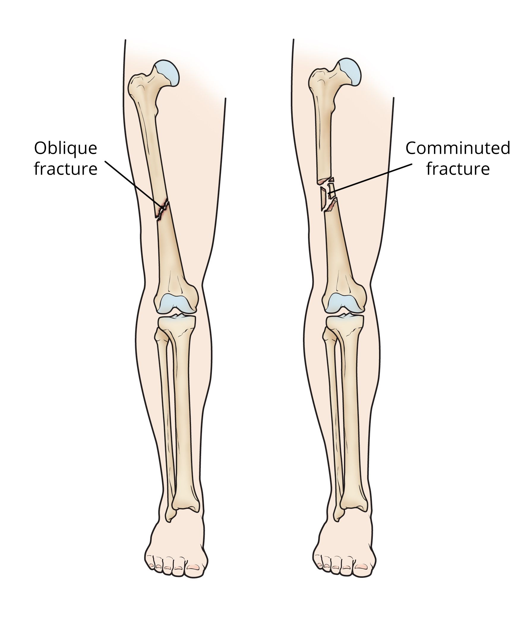
The specific mechanism of injury often dictates the type and location of the fracture. For example, a direct blow to the side of the thigh might cause a transverse or oblique fracture, while a rotational force could lead to a spiral fracture.
Recognizing the Symptoms of a Femur Fracture
The signs and symptoms of a femur fracture are usually dramatic and unmistakable due to the severity of the injury and the significant pain involved. Immediate medical attention is paramount upon suspecting such a fracture.
Immediate and Severe Pain
The hallmark symptom of a femur fracture is intense, excruciating pain in the thigh. This pain is often described as sharp and deep, and it is exacerbated by any attempt to move the leg or bear weight on it. The pain is a direct result of the broken bone fragments moving against each other and the surrounding tissues, as well as the inflammatory response.
Deformity of the Thigh
The injured leg may appear noticeably shorter than the uninjured leg. This shortening is due to the powerful muscles of the thigh contracting and pulling the broken bone fragments out of alignment. The thigh may also appear visibly angulated, bowed, or twisted. This deformity is a clear indicator of a significant disruption of the bone’s integrity.
Inability to Stand or Walk
Due to the severe pain and the loss of structural integrity, an individual with a femur fracture will be unable to bear any weight on the injured leg. Standing or attempting to walk will be impossible.
Swelling and Bruising
Significant swelling and bruising (ecchymosis) will typically develop around the fracture site. This is caused by bleeding from the fractured bone and surrounding soft tissues. The bruising may spread down the leg towards the knee or even into the calf due to gravity.
Open Fractures (Compound Fractures)
In some cases, a femur fracture can be an “open” or “compound” fracture, where the broken bone fragments pierce through the skin. This is a medical emergency due to the high risk of infection. The wound will be visible, and bone may be protruding from the skin.
It’s important to note that not all symptoms may be immediately apparent, and in some cases, the initial presentation might be less dramatic, especially in individuals with certain medical conditions. However, any suspicion of a femur fracture warrants immediate emergency medical evaluation.
Diagnostic Approaches and Medical Management
Diagnosing a femur fracture involves a combination of physical examination, imaging techniques, and a thorough medical history. Once diagnosed, the management focuses on stabilizing the fracture, promoting healing, and restoring function.
Diagnostic Tools
- Physical Examination: A healthcare professional will assess the injured leg for signs of deformity, swelling, bruising, and tenderness. They will also check for nerve and blood vessel damage by assessing sensation, pulses, and motor function in the foot and lower leg.
- X-rays: Standard X-rays are the primary imaging modality for diagnosing femur fractures. They provide clear images of the bone, allowing physicians to determine the location, type, and severity of the fracture. Multiple views are typically taken to get a comprehensive understanding of the fracture pattern.
- CT Scans (Computed Tomography): In complex fractures, particularly those involving the joint surfaces or with multiple fragments, a CT scan may be ordered. CT scans provide more detailed cross-sectional images of the bone, which can be crucial for surgical planning.
- MRI Scans (Magnetic Resonance Imaging): While not typically used for initial fracture diagnosis, an MRI might be employed to assess associated soft tissue injuries, such as ligament tears, muscle damage, or cartilage injuries, which are common in femur fractures.
Treatment Modalities
The treatment for a femur fracture is highly dependent on the location, type, and severity of the break, as well as the patient’s age, overall health, and activity level. Surgical intervention is almost always required for femur shaft fractures.
Non-Surgical Management (Rare for Femur Shaft Fractures)
Historically, traction was used to manage femur fractures, but this is rarely employed today as a primary treatment for adult femur shaft fractures due to complications and prolonged immobility. It may be used temporarily to stabilize the limb in certain pediatric cases or in preparation for surgery.
Surgical Management (The Standard of Care)
Surgery is the cornerstone of treatment for most femur fractures, aiming to realign the bone fragments and stabilize them to allow for healing.
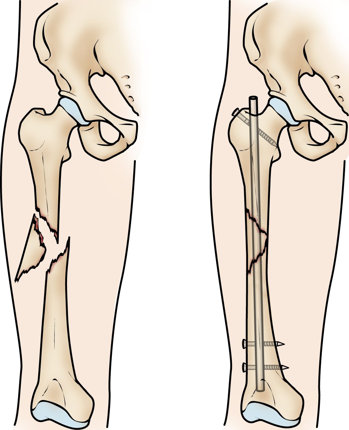
- Intramedullary Nailing: This is the most common surgical technique for femur shaft fractures. A metal rod (nail) is inserted down the hollow center of the femur, extending from the hip to just above the knee. Screws are then used to fix the nail to the bone at both ends, holding the fracture fragments in place. This method allows for early mobilization and weight-bearing, which is crucial for recovery.
- Plates and Screws: For certain types of fractures, particularly those closer to the hip or knee joint, plates and screws may be used. A metal plate is positioned along the outside of the bone, and screws are used to attach it to the bone fragments. This is often the preferred method for distal femur fractures.
- External Fixation: In cases of severe open fractures or when there is significant soft tissue damage, an external fixator may be used. This involves pins or screws inserted into the bone fragments above and below the fracture site, which are then connected to an external frame. This provides stability while allowing access to the wound for cleaning and dressing.
Rehabilitation and Recovery
Following treatment, a comprehensive rehabilitation program is essential for a successful recovery. This typically involves:
- Physical Therapy: Early mobilization and gradual progression of exercises are crucial. Physical therapists will guide patients through exercises to regain range of motion in the hip and knee, strengthen the surrounding muscles, and improve balance and gait.
- Pain Management: Effective pain management is vital throughout the recovery process, often involving medication and other therapeutic modalities.
- Weight-Bearing Progression: The ability to bear weight on the injured leg will be gradually introduced and increased under the guidance of the healthcare team.
- Return to Activity: The timeline for returning to normal activities and sports varies greatly depending on the severity of the fracture and the individual’s response to treatment and rehabilitation.
The Long-Term Outlook and Potential Complications
While femur fractures are serious injuries, advancements in surgical techniques and rehabilitation have significantly improved the prognosis for most patients. However, like any major orthopedic trauma, there are potential long-term implications and complications to consider.
Factors Influencing Recovery
The speed and completeness of recovery are influenced by several factors:
- Age: Younger patients generally heal faster and have a better capacity to regain full function compared to older adults.
- Overall Health: Pre-existing medical conditions, such as diabetes or cardiovascular disease, can affect healing and increase the risk of complications.
- Fracture Severity: Complex fractures with multiple fragments, open fractures, or those involving joint surfaces may require longer healing times and more intensive rehabilitation.
- Adherence to Rehabilitation: Diligent participation in physical therapy and following medical advice are paramount for optimal outcomes.
Potential Complications
Despite best efforts, certain complications can arise following a femur fracture:
- Infection: This is a particular concern with open fractures or those requiring surgical implants. Prompt treatment with antibiotics is crucial.
- Non-Union or Malunion: Non-union occurs when the bone fails to heal completely, while malunion happens when the bone heals in an incorrect position. These may require further surgical intervention.
- Blood Clots (Deep Vein Thrombosis – DVT): Immobility after a fracture increases the risk of blood clots forming in the leg veins. Anticoagulant medications and early mobilization help mitigate this risk.
- Nerve or Blood Vessel Damage: While surgeons strive to avoid these, accidental damage to surrounding nerves or blood vessels during the initial injury or surgery is a possibility, though relatively rare.
- Stiffness and Reduced Range of Motion: Scar tissue formation and prolonged immobility can lead to stiffness in the hip and knee joints.
- Post-Traumatic Arthritis: Fractures that extend into the knee or hip joint can increase the risk of developing arthritis in that joint later in life.
- Leg Length Discrepancy: If the fracture does not heal perfectly aligned, a slight difference in leg length can occur, which may require shoe lifts to correct.
- Pain: Chronic pain in the affected leg can persist in some individuals.
Long-Term Outlook
For many, the long-term outlook after a femur fracture is positive, with a return to most pre-injury activities possible. However, some individuals may experience persistent pain, limitations in mobility, or an increased susceptibility to future injuries in the affected limb. Regular follow-up with orthopedic specialists is recommended to monitor healing and address any emerging concerns. Educating oneself about the injury, actively participating in rehabilitation, and maintaining a healthy lifestyle are key to navigating the recovery process and achieving the best possible long-term outcome after a femur bone fracture.
aViewFromTheCave is a participant in the Amazon Services LLC Associates Program, an affiliate advertising program designed to provide a means for sites to earn advertising fees by advertising and linking to Amazon.com. Amazon, the Amazon logo, AmazonSupply, and the AmazonSupply logo are trademarks of Amazon.com, Inc. or its affiliates. As an Amazon Associate we earn affiliate commissions from qualifying purchases.