
The human body is a marvel of intricate biological engineering, and understanding its inner workings has been a relentless pursuit for humanity. In the realm of modern medicine, this pursuit has been profoundly amplified by technological advancements, none more impactful than medical imaging. Among the array of sophisticated diagnostic tools, the Computed Tomography (CT) scan, specifically a CT thorax, stands out as a cornerstone in visualizing and understanding the complex anatomy of the chest. But what exactly is a CT thorax, and what cutting-edge technology underpins this vital diagnostic procedure?
Beyond its immediate medical application, the principles and advancements in CT technology resonate with the broader themes of innovation we explore in the Tech world. From the intricate algorithms that process vast amounts of data to the sophisticated hardware that captures detailed images, CT scanning is a testament to human ingenuity. Furthermore, the branding of these sophisticated medical devices, the marketing strategies employed to introduce them to healthcare providers, and the ongoing development in AI-driven diagnostic tools all align with our Brand and Money sections, showcasing how innovation translates into tangible medical solutions and economic impact.
Understanding the Fundamentals: What is a CT Scan?
At its core, a Computed Tomography (CT) scan is a non-invasive medical imaging technique that utilizes X-rays to create detailed, cross-sectional images of the body. Unlike a conventional X-ray, which produces a single, two-dimensional image, a CT scanner takes multiple X-ray beams from different angles and combines them using a computer to generate highly detailed slices of anatomy. Think of it like slicing a loaf of bread; each slice provides a distinct view of the interior.
The technology behind this process is a sophisticated interplay of hardware and software. The CT scanner itself is a large, donut-shaped machine. Inside this ring, an X-ray tube rotates around the patient, emitting a focused beam of X-rays. As the X-rays pass through the body, they are attenuated (weakened) to varying degrees depending on the density of the tissues they encounter. Bones, for instance, absorb more X-rays than soft tissues or air.
The Physics of Imaging: How X-rays Paint a Picture
The attenuated X-ray beams are then detected by an array of sensitive detectors positioned on the opposite side of the X-ray tube. These detectors measure the intensity of the X-rays that have passed through the patient. This data, representing countless measurements from every angle, is then transmitted to a powerful computer.
Here’s where the “computed” in Computed Tomography comes into play. Sophisticated algorithms, often leveraging principles of mathematical reconstruction, are employed to process this raw data. These algorithms essentially work backward, using the attenuation information from all angles to reconstruct a precise, cross-sectional image of the body. The computer assigns different shades of gray to different tissue densities – white for dense bone, black for air, and various shades of gray for soft tissues like organs and muscles.
From Slices to 3D: The Power of Reconstruction
The output of a CT scanner is a series of axial (horizontal) slices. However, with advanced software, these individual slices can be digitally stacked and reconstructed to create three-dimensional (3D) images. This allows radiologists and clinicians to view the anatomy from various perspectives, providing a more comprehensive understanding of its structure and relationships. This capability is invaluable for surgical planning and for identifying complex abnormalities.
The speed and precision of modern CT scanners have dramatically improved over the years. Early CT scanners could take minutes to acquire a single slice, but today’s helical or spiral scanners can acquire multiple slices in a matter of seconds, significantly reducing scan times and minimizing motion artifacts. This speed is crucial for imaging dynamic organs like the heart or for patients who have difficulty remaining still.
Zooming In: The CT Thorax Explained
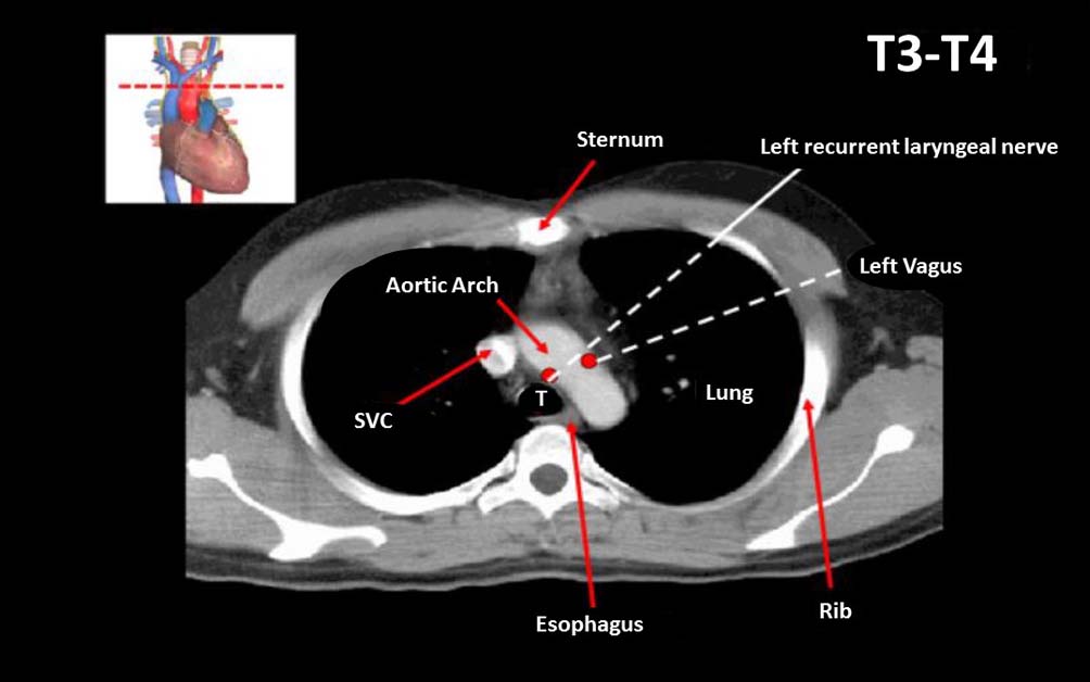
Now, let’s specifically address the “CT thorax.” This refers to a CT scan performed on the thoracic region of the body, which encompasses the chest. The thorax houses vital organs such as the lungs, heart, major blood vessels (aorta, pulmonary arteries), esophagus, and trachea, as well as the ribs, sternum, and spine.
A CT thorax is performed when a physician needs a detailed view of these structures, often to diagnose or monitor a wide range of conditions. The procedure is similar to a general CT scan: the patient lies on a table that moves through the center of the CT scanner. For a CT thorax, the patient is typically positioned on their back, with their arms raised above their head to avoid obstructing the X-ray beam.
Why a CT Thorax? Illuminating the Chest’s Interior
The indications for a CT thorax are diverse and critical in diagnosing various pathologies:
- Lung Diseases: This is perhaps the most common application. CT thorax excels at detecting and characterizing lung nodules (potential tumors), pneumonia, pulmonary embolism (blood clots in the lungs), emphysema, interstitial lung disease (scarring of the lung tissue), and other complex lung conditions that might not be fully visible on a standard chest X-ray.
- Heart and Blood Vessel Abnormalities: While specialized cardiac CT scans exist, a standard CT thorax can reveal abnormalities in the aorta, such as aneurysms (bulging of the aorta) or dissections (tears in the aortic wall). It can also help assess the extent of calcification in the coronary arteries.
- Trauma Assessment: In cases of chest trauma from accidents or injuries, a CT thorax can quickly identify fractures of the ribs or sternum, injuries to the lungs or heart, and internal bleeding.
- Cancer Staging and Monitoring: For patients diagnosed with cancer, a CT thorax is often used to determine the extent of the disease (staging) and to monitor the effectiveness of treatment by assessing tumor size and spread.
- Infections and Inflammatory Conditions: It can help diagnose and manage infections in the chest, such as empyema (pus in the chest cavity) or mediastinitis (inflammation of the mediastinum, the space between the lungs).
Contrast Agents: Enhancing Visibility
In many CT thorax examinations, a contrast agent is administered to further enhance the visualization of certain structures. This contrast agent is typically a liquid containing iodine, which appears opaque on X-rays. It can be administered intravenously (through a vein), orally, or rectally, depending on the area being examined.
For a CT thorax, intravenous contrast is commonly used. It helps to highlight blood vessels, making them clearly visible and allowing for the detection of blockages, aneurysms, or inflammation. It also helps to differentiate between different types of tissues and can improve the detection of tumors or areas of infection. The use of contrast agents is carefully considered based on the patient’s medical history, including any allergies or kidney issues.

The Technological Edge: Innovations Driving CT Imaging
The evolution of CT technology has been a continuous journey of innovation, deeply intertwined with advancements in computing, electronics, and materials science. These developments not only enhance image quality and diagnostic accuracy but also improve patient safety and workflow efficiency.
Advancements in Detector Technology: Capturing More Data, Faster
The detectors in a CT scanner are crucial for accurately measuring the attenuated X-ray beams. Modern detectors are highly sensitive and can capture X-ray photons with remarkable efficiency. This has led to the development of multi-detector CT (MDCT) scanners, which feature multiple rows of detectors arranged in a helical pattern. MDCT scanners can acquire data much faster, allowing for thinner slices and shorter scan times. This translates to improved spatial resolution (the ability to distinguish small details) and reduced patient exposure to radiation.
Iterative Reconstruction and AI: Sharpening Images, Reducing Dose
Historically, CT image reconstruction relied heavily on analytical methods like filtered back-projection. However, more recent innovations have introduced iterative reconstruction techniques. These methods involve repeatedly refining the image based on mathematical models and comparing the reconstructed image back to the raw data. This process allows for the generation of high-quality images with significantly lower radiation doses compared to older methods.
The integration of Artificial Intelligence (AI) is further revolutionizing CT imaging. AI algorithms are being developed for a multitude of applications, including:
- Image Denoising: Reducing image noise, which can obscure subtle details, leading to clearer images.
- Image Segmentation: Automatically identifying and outlining specific organs or lesions, aiding in quantitative analysis and measurement.
- Automated Detection: Flagging potential abnormalities, such as lung nodules or pulmonary embolisms, for radiologists to review.
- Dose Optimization: Dynamically adjusting radiation parameters to achieve diagnostic quality images with the lowest possible radiation dose for each individual patient.
These AI-powered tools not only improve diagnostic accuracy and efficiency but also contribute to a more personalized approach to medical imaging. The ongoing research and development in AI for medical imaging strongly echo the rapid advancements we see in the broader Tech landscape.
Dose Reduction Strategies: Balancing Risk and Reward
Patient safety is paramount in medical imaging. CT scans, while invaluable, do involve exposure to ionizing radiation. Therefore, significant efforts have been made to reduce radiation doses without compromising diagnostic quality. This includes:
- Automatic Exposure Control (AEC): Adjusting the X-ray beam intensity based on the patient’s size and the area being scanned.
- Low-Dose Protocols: Employing specialized imaging protocols designed for reduced radiation exposure, often utilizing iterative reconstruction techniques.
- Photon-Counting Detectors: A newer generation of detectors that can directly count X-ray photons, offering improved spectral information and the potential for even lower doses and enhanced image quality.
The continuous pursuit of lower radiation doses while maintaining or improving image quality reflects a broader trend in technology design – optimizing performance while minimizing negative impacts. This dedication to responsible innovation is a hallmark of progress across all fields.
The Broader Impact: From Medical Marvel to Technological Showcase
The CT thorax, while a specialized medical procedure, represents a culmination of technological advancements that resonate with several key themes. The development and deployment of such sophisticated imaging systems involve significant investment, intricate engineering, and strategic marketing – all areas we explore within our Money and Brand sections.
Branding and Marketing Advanced Medical Technology
The manufacturers of CT scanners are global players who invest heavily in research and development, aiming to create the most advanced, reliable, and user-friendly machines. The Brand of a medical imaging company is built on reputation for innovation, accuracy, and patient care. This involves extensive marketing efforts, showcasing their technology at medical conferences, publishing case studies, and developing strong relationships with healthcare institutions. The development of a compelling corporate identity is crucial for establishing trust and credibility in a highly regulated and competitive industry.
The Financial Ecosystem of Medical Imaging
The cost of developing, manufacturing, and maintaining CT scanners is substantial. This impacts the healthcare economy, influencing the pricing of procedures and the capital expenditure of hospitals. The Money aspect extends to the financial tools and strategies employed by these companies for funding research, managing production, and generating revenue. Furthermore, the increasing use of AI in CT interpretation can lead to new business models and investment opportunities, as companies develop specialized software solutions and platforms. The efficiency gains and potential for improved patient outcomes driven by these technologies can also translate into significant cost savings for healthcare systems in the long run.

The Future: AI-Powered Diagnostics and Personalized Medicine
The trajectory of CT technology, particularly with the integration of AI, points towards a future of increasingly personalized and predictive medicine. AI can help identify subtle patterns that the human eye might miss, leading to earlier diagnosis and more tailored treatment plans. This push towards greater precision and individualization in healthcare is a powerful demonstration of how technology, when applied thoughtfully, can profoundly improve human well-being.
In conclusion, a CT thorax is a sophisticated medical imaging technique that provides incredibly detailed insights into the chest cavity. Behind this vital diagnostic tool lies a rich tapestry of technological innovation, from the fundamental physics of X-ray attenuation to the cutting-edge advancements in detectors, reconstruction algorithms, and artificial intelligence. Understanding what a CT thorax is goes beyond just its medical function; it’s a glimpse into the powerful synergy between science, engineering, and human ingenuity that continues to shape the future of healthcare and beyond.
aViewFromTheCave is a participant in the Amazon Services LLC Associates Program, an affiliate advertising program designed to provide a means for sites to earn advertising fees by advertising and linking to Amazon.com. Amazon, the Amazon logo, AmazonSupply, and the AmazonSupply logo are trademarks of Amazon.com, Inc. or its affiliates. As an Amazon Associate we earn affiliate commissions from qualifying purchases.