The human head, a marvel of biological engineering, houses our consciousness, our senses, and the intricate network that defines who we are. When concerns arise regarding its health, medical imaging becomes a crucial diagnostic tool. Among these, the Computed Tomography (CT) scan of the head stands out as a powerful technological advancement that offers invaluable insights. But what exactly is this procedure, and how does it leverage cutting-edge technology to aid in understanding the complexities of the brain and its surrounding structures? This article will delve into the world of head CT scans, exploring their technological underpinnings, their application in various medical scenarios, and how understanding this technology can empower individuals to make more informed decisions about their health, resonating with the core interests of tech, brand, and money-related insights found on this platform.
The Technological Heartbeat: How a Head CT Scan Works
At its core, a CT scan of the head is a sophisticated imaging technique that utilizes X-rays and advanced computational processing to create detailed cross-sectional images, or “slices,” of the brain and surrounding bony structures. This stands in stark contrast to a traditional X-ray, which produces a single, flat image. The technology behind a CT scanner is a testament to decades of innovation in physics, computer science, and engineering.
The Mechanics of Imaging: X-rays and Detectors
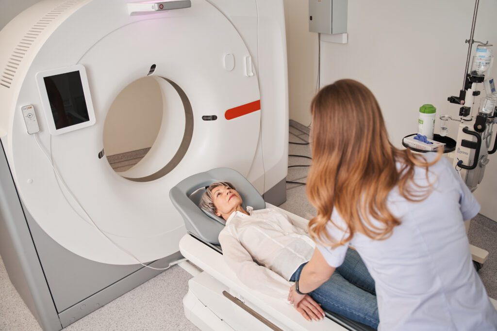
The CT scanner itself is a doughnut-shaped machine that encases a rotating X-ray tube and a bank of sensitive X-ray detectors. As the patient lies on a motorized table that slides through the opening of the scanner, the X-ray tube rapidly circles the head, emitting a thin, fan-shaped beam of X-rays. These X-rays pass through the different tissues of the head – bone, soft tissue, fluid, and air – each absorbing the radiation to a different degree. Denser materials, like bone, absorb more X-rays, while less dense materials, like air, absorb fewer.
The X-ray detectors, positioned opposite the X-ray tube, capture the attenuated X-ray beams that have passed through the body. These detectors measure the intensity of the X-rays that reach them, converting this information into electronic signals. The rapid rotation of the X-ray tube and detectors allows for multiple projections of the head to be taken from various angles in a very short period. This comprehensive data acquisition is critical for the subsequent image reconstruction.
The Digital Alchemy: Reconstruction and Visualization
The raw data collected by the detectors is not an image in itself. It’s a series of measurements representing the X-ray absorption at countless points from multiple angles. This is where the “computed” aspect of Computed Tomography comes into play. Powerful computer algorithms, employing complex mathematical techniques like the filtered back-projection or iterative reconstruction algorithms, process this vast amount of data.
These algorithms essentially “reconstruct” the internal structures of the head from the multiple X-ray projections. They analyze how the X-rays were absorbed from each angle to determine the density of each point within the scanned volume. The result is a series of highly detailed cross-sectional images, often referred to as “slices.” These slices can be viewed individually, akin to looking at slices of a loaf of bread, or they can be digitally stacked and processed to create three-dimensional (3D) reconstructions.
Modern CT scanners are remarkably fast, capable of acquiring these detailed slices in mere seconds. This speed is crucial, especially in emergency situations where time is of the essence, such as after a head injury. Furthermore, advancements in detector technology have led to multi-detector CT (MDCT) scanners, which have multiple rows of detectors. This allows for the acquisition of thinner slices and faster scanning, providing even greater detail and reducing motion artifacts.
The Role of Contrast Agents
In some instances, a contrast agent, typically an iodine-based solution, may be administered intravenously before or during the scan. This agent is designed to highlight specific structures, such as blood vessels or tumors, by altering their X-ray attenuation. When the contrast agent flows through the blood vessels, it makes them appear brighter on the CT images, allowing radiologists to better assess blood flow, identify blockages, or detect abnormalities within the vascular system of the brain. The decision to use contrast is based on the suspected medical condition and is a critical component of optimizing diagnostic accuracy.
Illuminating the Intricacies: Applications of Head CT Scans
The detailed anatomical information provided by a head CT scan makes it an indispensable tool for diagnosing and monitoring a wide range of neurological conditions. Its ability to quickly visualize bone, blood, and soft tissue makes it particularly valuable in emergency settings, but its utility extends far beyond acute trauma.
Emergency Diagnostics: Responding to Critical Events
One of the most common and critical applications of a head CT scan is in the evaluation of head trauma. Following an accident, fall, or any impact to the head, a CT scan can rapidly identify the presence of:
- Intracranial Hemorrhage: This includes bleeding within the brain itself (intraparenchymal hemorrhage), between the brain and its outer membrane (subdural hematoma), or between the skull and the outer membrane (epidural hematoma). These are life-threatening conditions that require immediate medical intervention.
- Skull Fractures: CT scans are highly sensitive in detecting even hairline fractures of the skull, which can be crucial for determining the severity of an injury and the need for further management.
- Cerebral Edema: Swelling of the brain tissue, which can occur after trauma, can also be visualized on CT scans.

Beyond trauma, CT scans of the head are vital in diagnosing and managing strokes. A non-contrast CT scan is often the first imaging modality used to rule out a hemorrhagic stroke (bleeding in the brain). If no blood is detected, but stroke symptoms are present, a contrast-enhanced CT angiography (CTA) may be performed to assess for an ischemic stroke (blockage of blood flow) by visualizing the blood vessels.
Unraveling Neurological Disorders: Beyond Acute Crises
The diagnostic power of head CT scans extends to a variety of non-emergency neurological conditions:
- Tumors: CT scans can detect and characterize brain tumors, differentiating between solid masses and fluid-filled cysts. While MRI is often preferred for detailed evaluation of soft tissue, CT plays a crucial role in initial detection, particularly in emergency situations or when MRI is contraindicated.
- Infections: Abscesses or other signs of infection within the brain can be identified.
- Congenital Abnormalities: Some structural abnormalities present from birth can be visualized.
- Degenerative Diseases: While MRI is generally superior for visualizing the subtle changes associated with neurodegenerative conditions like Alzheimer’s or Parkinson’s disease, CT can sometimes reveal significant atrophy or other gross structural changes.
- Monitoring Treatment: CT scans are used to monitor the effectiveness of treatments for various neurological conditions, such as tracking the size of a tumor after radiation therapy or assessing the extent of recovery after a stroke.
The “Brand” of Health Decisions: Empowering Patients Through Technological Understanding
Understanding the technology behind a head CT scan, even at a foundational level, can significantly empower individuals when facing medical decisions. This knowledge contributes to a more informed “brand” of patient engagement, fostering better communication with healthcare providers and a greater sense of control over one’s health journey.
Navigating the Healthcare Landscape with Informed Choices
When a doctor recommends a CT scan of the head, having a basic understanding of what it entails can alleviate anxiety and facilitate a more productive discussion. Knowing that it involves X-rays and computer processing, and that it can detect a range of abnormalities, helps demystify the procedure. This knowledge can also help patients ask more pertinent questions, such as:
- “What specifically are we looking for with this scan?”
- “Is a contrast agent necessary, and what are the potential risks?”
- “How quickly will I receive the results, and what are the next steps?”
This proactive approach to healthcare aligns with building a strong personal health “brand” – one that is informed, engaged, and collaborative with medical professionals.
The Cost-Benefit Equation: Understanding the “Money” Aspect
In the realm of healthcare, understanding the financial implications of diagnostic procedures is also crucial. CT scans, while invaluable, represent a significant investment in healthcare resources.
- Direct Costs: The cost of a CT scan can vary widely depending on the facility, geographic location, and whether contrast is used. This cost is typically covered by health insurance, but understanding deductibles, co-pays, and out-of-pocket maximums is essential for personal financial planning.
- Indirect Benefits: The “money” saved or gained through a timely and accurate diagnosis from a CT scan can be substantial. Early detection of a stroke, for instance, can lead to more effective treatment, potentially reducing long-term disability and associated healthcare costs, as well as enabling a quicker return to work or daily activities. Similarly, identifying a treatable brain tumor early can significantly improve prognosis and reduce the overall financial burden of care.
- Technological Advancements and Cost-Effectiveness: The ongoing advancements in CT technology, while often leading to initial higher costs for state-of-the-art equipment, also aim to improve efficiency and reduce radiation exposure. As technology matures and becomes more widespread, the cost-effectiveness of CT scans generally improves, making this vital diagnostic tool more accessible.

The Future of Head Imaging: Integration and Innovation
The field of medical imaging is constantly evolving, with AI and machine learning playing an increasingly significant role. We are already seeing AI being used to:
- Enhance Image Quality: Algorithms can reduce noise and artifacts in CT scans, leading to clearer images.
- Automate Analysis: AI can assist radiologists in identifying potential abnormalities, flagging areas of concern for closer review.
- Predict Outcomes: Future applications may involve using AI to predict the likelihood of certain outcomes based on CT scan findings.
These technological strides promise to make head CT scans even more accurate, efficient, and potentially more affordable in the long run. Staying abreast of these developments not only enhances our understanding of current medical practices but also prepares us for the future of healthcare, where technology and personalized medicine will be inextricably linked.
In conclusion, a CT scan of the head is a powerful diagnostic tool that leverages sophisticated X-ray technology and advanced computational processing to create detailed images of the brain. Understanding its mechanics, its diverse applications, and its implications for personal health decisions, financial planning, and the broader landscape of technological innovation allows individuals to approach their health with greater confidence and informed agency. By bridging the gap between complex technology and everyday health concerns, we can foster a more empowered and proactive approach to well-being.
aViewFromTheCave is a participant in the Amazon Services LLC Associates Program, an affiliate advertising program designed to provide a means for sites to earn advertising fees by advertising and linking to Amazon.com. Amazon, the Amazon logo, AmazonSupply, and the AmazonSupply logo are trademarks of Amazon.com, Inc. or its affiliates. As an Amazon Associate we earn affiliate commissions from qualifying purchases.