
In the intricate landscape of modern medicine, few technologies offer the profound diagnostic clarity and non-invasive insight of Magnetic Resonance Imaging (MRI). Far more than just another medical scan, MRI stands as a pinnacle of technological ingenuity, employing sophisticated physics and advanced computing to peer deep within the human body. It reveals structures and pathologies with unparalleled detail, transforming how medical professionals diagnose, monitor, and treat a vast array of conditions. From the delicate neural pathways of the brain to the complex architecture of joints and soft tissues, an MRI scan shows a world invisible to the naked eye, offering a silent, radiant window into our biological machinery.
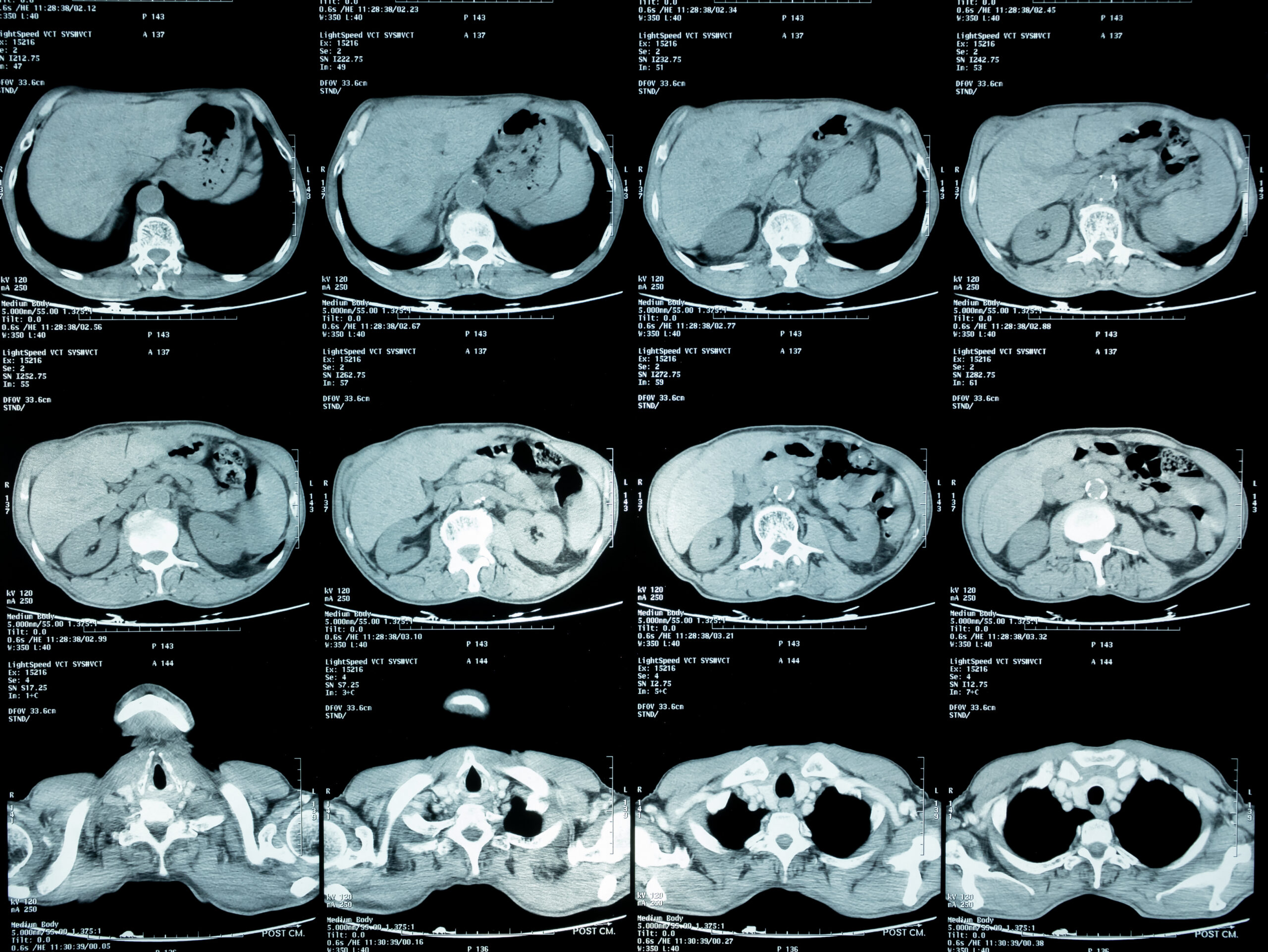
At its core, MRI is a triumph of engineering, data science, and biomedical application. It represents a leap beyond conventional imaging techniques like X-rays and CT scans, particularly in its ability to differentiate between various soft tissues – a critical advantage when evaluating organs, muscles, ligaments, and the central nervous system. This article delves into the technological underpinnings of MRI, exploring how this remarkable system works, the comprehensive view it offers across different body systems, its cutting-edge applications, and the considerations that shape its use in contemporary healthcare.
The Technological Marvel Behind MRI: How It Works
Understanding what an MRI scan shows begins with appreciating the intricate technology that powers it. Unlike X-rays, which use ionizing radiation, or ultrasound, which uses sound waves, MRI leverages the fundamental properties of atomic nuclei within the body, specifically hydrogen atoms. This difference is not merely academic; it’s the foundation of MRI’s safety and exceptional image quality for soft tissues.
Beyond X-rays: The Physics of MRI
The core principle of MRI revolves around powerful superconducting magnets, radiofrequency coils, and the ubiquitous presence of hydrogen atoms in water molecules throughout the body. When a patient enters an MRI scanner, the powerful magnetic field (measured in Teslas, typically 1.5T or 3T for clinical use) aligns the protons (which are essentially the nuclei of hydrogen atoms) within the body’s water molecules. These protons normally spin in random directions, but under the influence of the magnet, they align either parallel or anti-parallel to the main magnetic field, much like tiny compass needles.
Next, a radiofrequency current is briefly pulsed through the patient. This pulse temporarily knocks the aligned protons out of alignment. When the radiofrequency pulse is turned off, the protons relax back into alignment with the main magnetic field. As they do, they release energy in the form of radio signals. The crucial insight here is that different tissues relax at different rates and emit signals of varying strengths, depending on their water content, fat content, and other molecular characteristics. This subtle variation is the secret to MRI’s incredible differentiation capabilities.
From Signal to Image: Data Acquisition and Processing
The emitted radio signals are detected by receiver coils within the MRI scanner. These raw signals are then fed into a sophisticated computer system, which employs complex mathematical algorithms and Fourier transforms to process the data. Critically, the MRI scanner also uses gradient coils, which create secondary, varying magnetic fields. These gradients allow the system to precisely pinpoint the origin of each signal, essentially giving the computer a 3D map of where each proton is located and how it’s behaving.
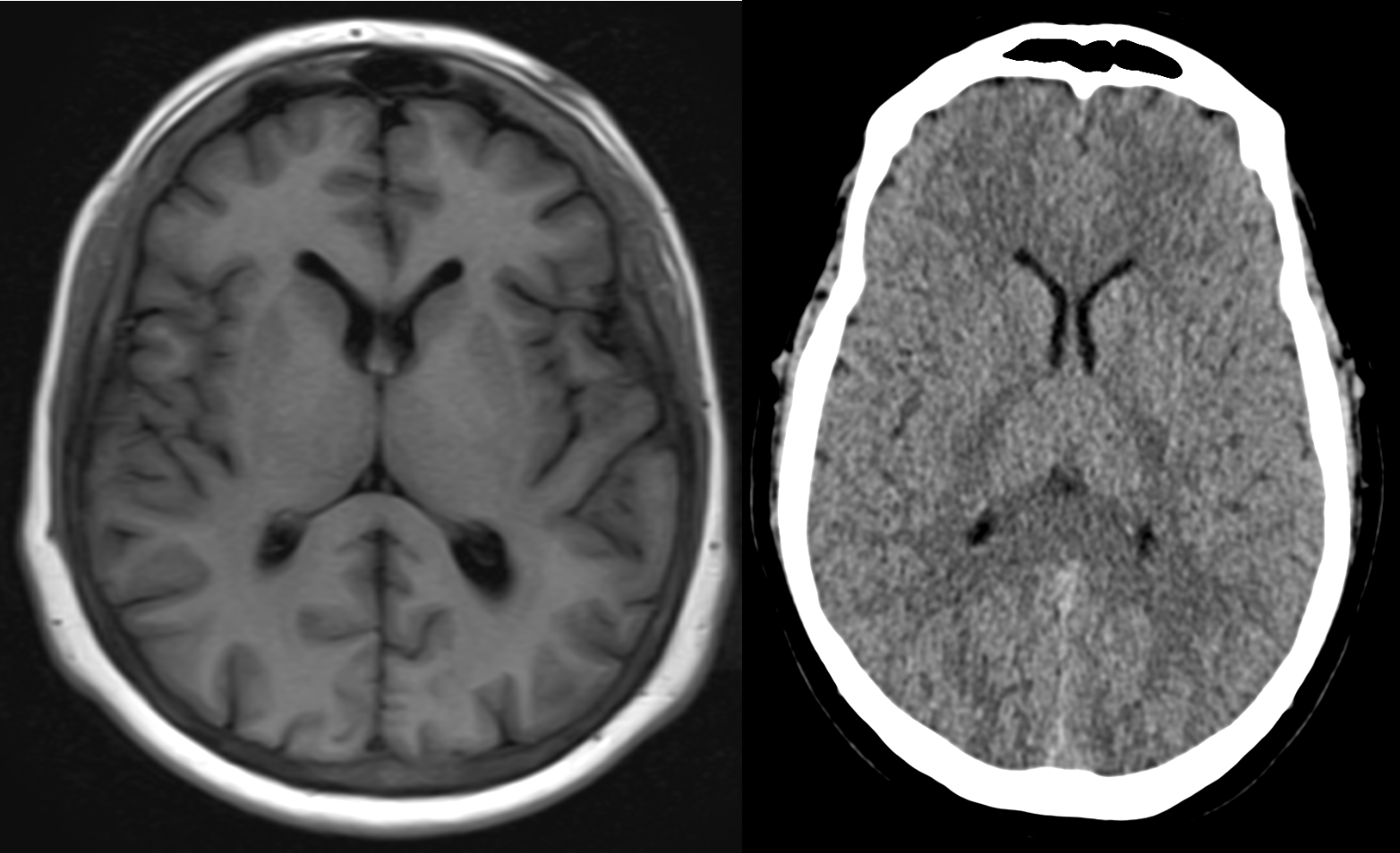
The computer then constructs detailed cross-sectional images or “slices” of the body. These images can be viewed in multiple planes (axial, sagittal, coronal), offering a comprehensive three-dimensional perspective. Radiologists can manipulate these images, applying different “sequences” (specific timing of radiofrequency pulses and signal detection) to highlight different tissue characteristics. For example, T1-weighted sequences are excellent for anatomical detail, while T2-weighted sequences are sensitive to water and inflammation. This flexibility in data acquisition and processing is a hallmark of MRI’s technological sophistication, allowing for tailored diagnostic approaches depending on the clinical question.
The Non-Invasive Advantage: Safety and Comfort
One of the most significant technological advantages of MRI is its non-invasive nature and the absence of ionizing radiation. This makes it a safer option for repeated scans, particularly for children and pregnant women, when compared to X-rays or CT scans. While the powerful magnetic field does require strict adherence to safety protocols regarding metallic implants (pacemakers, certain clips), the technology itself poses no direct radiation risk.
Improvements in scanner design, such as wider bores and “open MRI” configurations, have also addressed patient comfort issues like claustrophobia. Furthermore, the development of various contrast agents, often gadolinium-based, can enhance the visibility of certain tissues or abnormalities, allowing for even greater diagnostic precision. These agents are safely administered intravenously and aid in highlighting blood vessels, tumors, and areas of inflammation by altering the magnetic properties of surrounding tissues.
A Comprehensive View: What MRI Reveals Across Body Systems
The power of MRI lies in its versatility. Its ability to create high-contrast images of soft tissues makes it indispensable across almost every medical specialty. What an MRI scan shows is a detailed structural and, in some cases, functional map of various body systems, providing critical information for diagnosis and treatment planning.
Brain and Spinal Cord: Neurological Insights
For the central nervous system, MRI is the gold standard. It reveals an astonishing level of detail in the brain and spinal cord, making it invaluable for diagnosing:
-
Tumors: Distinguishing between benign and malignant growths, and assessing their size and location.
-
Strokes: Identifying areas of brain damage from ischemic or hemorrhagic events, even in early stages.
-
Multiple Sclerosis (MS): Detecting plaques (lesions) in the brain and spinal cord, crucial for diagnosis and monitoring disease progression.
-
Aneurysms and Vascular Malformations: Visualizing blood vessel abnormalities with MR angiography.
-
Spinal Cord Injuries and Disc Herniations: Precisely locating nerve compression, disc protrusions, and spinal cord damage.
-
Infections and Inflammatory Diseases: Identifying areas of infection or inflammation within neurological structures.
The superior soft tissue contrast of MRI allows neurologists and neurosurgeons to visualize the intricate structures of the brain – gray matter, white matter, ventricles, and the brainstem – with clarity unmatched by other imaging modalities.
Musculoskeletal System: Joints, Bones, and Soft Tissues
When it comes to the intricate network of bones, joints, muscles, ligaments, and tendons, MRI provides unparalleled diagnostic information. What an MRI scan shows here is crucial for orthopedic specialists:
-
Ligament and Tendon Tears: Such as ACL tears in the knee, rotator cuff tears in the shoulder, or Achilles tendon ruptures. MRI can accurately assess the extent and severity of these injuries.
-
Cartilage Damage: Detecting conditions like meniscal tears in the knee or articular cartilage defects in any joint.
-
Bone Marrow Abnormalities: Identifying stress fractures, infections (osteomyelitis), and tumors within the bone marrow, which might not be visible on X-rays.
-
Soft Tissue Tumors and Infections: Localizing and characterizing masses or areas of inflammation in muscles and other soft tissues.
-
Complex Joint Issues: Evaluating chronic pain, arthritis, and post-surgical complications with high precision.
For athletes and individuals with joint pain, MRI is often the decisive tool for pinpointing the exact cause of discomfort and guiding treatment, from physical therapy to surgical intervention.
Abdomen and Pelvis: Organs and Disease Detection
MRI offers a powerful, radiation-free way to examine the organs and structures within the abdomen and pelvis. What an MRI scan shows in this region includes:
-
Liver, Kidneys, Pancreas, and Spleen: Detecting tumors, cysts, inflammation, and evaluating organ function. For instance, MRI can differentiate between various types of liver lesions.
-
Gallbladder and Bile Ducts: Using MR cholangiopancreatography (MRCP) to visualize blockages or abnormalities without invasive procedures.
-
Reproductive Organs: Assessing conditions in the uterus, ovaries, and prostate gland, such as fibroids, endometriosis, cysts, and tumors.
-
Bowel Disease: Evaluating inflammatory bowel disease (Crohn’s disease, ulcerative colitis) for inflammation, strictures, and fistulas.
-
Adrenal Glands: Detecting tumors or hyperplasia.
MRI’s ability to provide excellent contrast between different soft tissues is particularly beneficial in the abdomen and pelvis, where many organs are densely packed and share similar densities with other imaging methods.
Cardiovascular System: Heart and Blood Vessels
Cardiac MRI (CMR) and MR Angiography (MRA) have revolutionized the assessment of the heart and blood vessels, offering detailed anatomical and functional information. What an MRI scan shows for the cardiovascular system includes:
-
Heart Muscle Damage: Identifying areas of scar tissue (e.g., after a heart attack), inflammation (myocarditis), and assessing viability.
-
Heart Function: Measuring ejection fraction, blood flow velocities, and wall motion, providing critical data on the heart’s pumping efficiency.
-
Congenital Heart Defects: Precisely visualizing complex anatomical abnormalities present from birth.
-
Blood Vessel Abnormalities: MRA can detect aneurysms, dissections, blockages (stenosis), and other vascular diseases in arteries throughout the body, including the brain, neck, chest, abdomen, and limbs, often without the need for iodinated contrast or radiation.
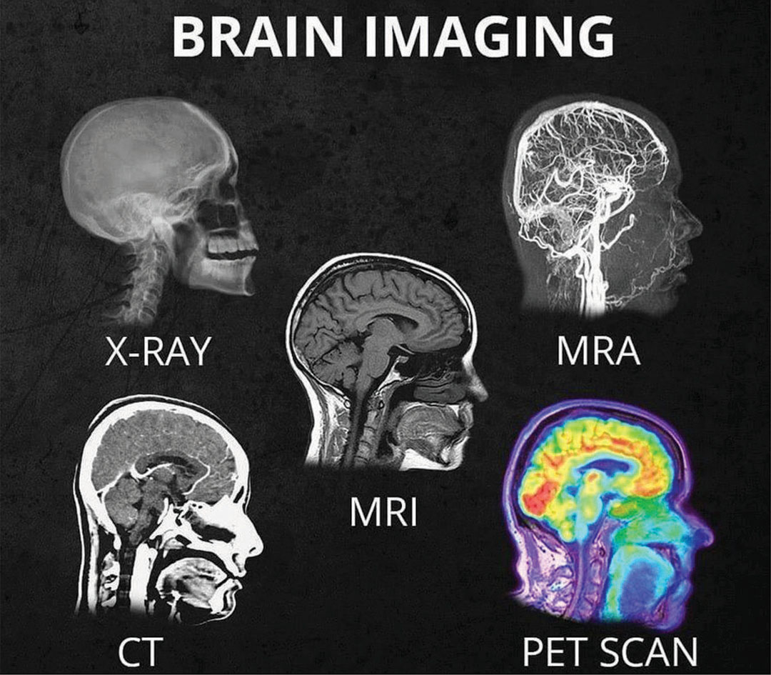
This non-invasive assessment of the cardiovascular system is invaluable for diagnosing complex conditions and guiding interventions, offering a safer alternative to more invasive angiographic procedures.
Advanced Applications and Emerging Trends in MRI Technology
The technological evolution of MRI is ongoing, pushing the boundaries of what these scans can reveal. Beyond standard anatomical imaging, advanced applications are providing unprecedented insights into function and microstructure, while new trends promise to further enhance diagnostic capabilities.
Functional MRI (fMRI): Mapping Brain Activity
Functional MRI (fMRI) is a cutting-edge application that measures brain activity by detecting changes associated with blood flow. When an area of the brain is more active, it demands more oxygenated blood. fMRI exploits the different magnetic properties of oxygenated and deoxygenated hemoglobin (the blood oxygenation level-dependent, or BOLD, contrast) to create maps of brain activity. What an fMRI scan shows are the specific regions of the brain that “light up” in response to tasks (e.g., moving a hand, speaking, listening, thinking).
This technology is primarily a research tool, used to understand cognitive processes, map sensory and motor areas, and investigate neurological and psychiatric disorders. Clinically, fMRI is sometimes used pre-surgically to map critical brain functions near tumors or lesions, helping neurosurgeons plan procedures to minimize damage to vital areas.
Diffusion Tensor Imaging (DTI): Visualizing Neural Pathways
Diffusion Tensor Imaging (DTI) is another specialized MRI technique that goes beyond anatomical structure to visualize the brain’s white matter tracts – the bundles of nerve fibers that connect different brain regions. DTI measures the microscopic movement (diffusion) of water molecules within these tissues. Because water molecules tend to diffuse more freely along the direction of nerve fibers rather than across them, DTI can infer the orientation and integrity of these pathways.
What a DTI scan shows is a detailed map of the brain’s “wiring,” often represented as colorful 3D images of fiber tracts (tractography). This is invaluable for:
-
Understanding Neurological Disorders: Investigating conditions like Alzheimer’s disease, Parkinson’s disease, and multiple sclerosis, where white matter integrity is affected.
-
Traumatic Brain Injury (TBI): Detecting subtle damage to white matter that might not be visible on conventional MRI.
-
Neurodevelopmental Disorders: Studying conditions like autism and ADHD by examining differences in brain connectivity.
AI and Machine Learning in MRI: Enhancing Diagnostics
The integration of Artificial Intelligence (AI) and Machine Learning (ML) is rapidly transforming MRI technology, promising to make scans faster, more accurate, and more accessible. What AI in MRI shows is a pathway to optimized workflows and improved diagnostic yield:
-
Accelerated Acquisition: AI algorithms can reconstruct high-quality images from less raw data, significantly reducing scan times, which is beneficial for patient comfort and throughput.
-
Noise Reduction and Image Enhancement: ML models can effectively denoise images and improve resolution, leading to clearer diagnostic pictures.
-
Automated Analysis: AI can assist in the automated segmentation of organs, measurement of volumes, and even the detection of subtle abnormalities (e.g., small tumors or lesions), potentially reducing radiologist workload and inter-observer variability.
-
Personalized Imaging: AI could help tailor scanning protocols to individual patient needs, optimizing image quality and diagnostic information for specific clinical questions.
This synergy between advanced imaging physics and computational intelligence represents a significant leap forward in diagnostic medical technology.
The Future of MRI: Ultra-High Field Magnets and Portable Systems
The future of MRI technology is characterized by a dual pursuit: greater detail and increased accessibility.
-
Ultra-High Field Magnets: Research is actively progressing with MRI scanners featuring ultra-high magnetic field strengths (e.g., 7 Tesla, 11 Tesla, and even higher). What these advanced systems show is even finer anatomical detail and more precise functional insights, particularly for the brain and small structures. These are primarily research tools for now but hold immense potential for future clinical applications.
-
Portable and Point-of-Care Systems: At the other end of the spectrum, efforts are underway to develop smaller, more portable, and lower-field MRI systems. The goal is to make MRI more accessible in emergency rooms, rural clinics, or even intensive care units, reducing the need to transport critically ill patients and expanding diagnostic capabilities beyond large medical centers. These systems may not offer the same exquisite detail as high-field scanners, but they promise to fill a critical gap in rapid, on-site imaging.
Limitations and Considerations: The Other Side of the Technological Coin
While MRI is an unparalleled diagnostic tool, it’s essential to acknowledge its limitations and the practical considerations that shape its deployment and utility within the healthcare system. These are not failures of the technology but rather inherent challenges that medical professionals and engineers continuously work to mitigate.
Challenges of Compatibility and Claustrophobia
The powerful magnetic field of an MRI scanner necessitates strict screening for metallic objects. What an MRI scan cannot show (or safely perform for) are individuals with certain metallic implants like pacemakers, certain types of aneurysm clips, cochlear implants, or some metallic foreign bodies, as these can be hazardous or interfere with image quality. While many modern implants are now “MRI-compatible,” careful patient screening remains paramount.
Furthermore, the enclosed space and loud noise generated by the gradient coils can induce claustrophobia and anxiety in some patients. Technological solutions like wider bore scanners, open MRI systems, and advanced noise reduction techniques help, but some patients may still require sedation. What an MRI scan shows is contingent on patient cooperation and stillness, which can be challenging for those uncomfortable within the scanner.
Cost and Accessibility: Bridging the Gap
MRI technology is expensive. The initial investment in the scanner itself, along with the high operational costs (maintenance, specialized staff, power consumption), makes MRI scans relatively costly for patients and healthcare systems. This often impacts accessibility, particularly in underserved regions or countries with limited healthcare budgets.
What an MRI scan shows is invaluable, but the economic barrier can mean that not everyone who needs one can readily access it. Efforts to improve efficiency (faster scan times, greater patient throughput), develop more affordable systems, and optimize reimbursement policies are ongoing to bridge this accessibility gap.
Interpreting the Data: The Human Element in Tech
Despite the technological sophistication of MRI, the interpretation of the complex images it produces remains a highly specialized skill. What an MRI scan shows is raw data – patterns, shades, and textures – that require the expertise of a trained radiologist. Radiologists undergo years of specialized training to accurately identify subtle abnormalities, differentiate between various pathologies, and correlate findings with clinical symptoms.
Even with the rise of AI in medical imaging, the human element of critical thinking, clinical context integration, and nuanced judgment remains indispensable. AI tools can assist, but the final diagnostic pronouncement and its integration into a patient’s care plan continue to rely on the professional insight of medical experts.
Conclusion
What does an MRI scan show? It reveals a meticulously detailed interior landscape of the human body, offering diagnostic insights that were once unimaginable. From its fundamental physics employing powerful magnets and radio waves to its advanced applications in functional imaging and AI integration, MRI stands as a testament to humanity’s drive to understand and heal. It is a technological marvel that has revolutionized neurological, musculoskeletal, abdominal, and cardiovascular diagnostics, becoming an indispensable tool in the modern medical arsenal.
As the technology continues to evolve with ultra-high field systems and increasing AI integration, MRI promises even greater precision and accessibility. While challenges remain concerning cost, compatibility, and patient comfort, ongoing innovation is steadily addressing these. Ultimately, the ability of an MRI scan to provide a safe, non-invasive, and remarkably clear window into the body’s most intricate structures solidifies its position as a cornerstone of medical technology, continuing to empower clinicians and improve patient outcomes worldwide.
aViewFromTheCave is a participant in the Amazon Services LLC Associates Program, an affiliate advertising program designed to provide a means for sites to earn advertising fees by advertising and linking to Amazon.com. Amazon, the Amazon logo, AmazonSupply, and the AmazonSupply logo are trademarks of Amazon.com, Inc. or its affiliates. As an Amazon Associate we earn affiliate commissions from qualifying purchases.