
Retinitis pigmentosa (RP) represents a complex, genetically heterogeneous group of inherited disorders that primarily affect the retina, the light-sensitive tissue lining the back of the eye. It is characterized by the progressive degeneration of photoreceptor cells—the rods and cones responsible for converting light into electrical signals sent to the brain. Because RP is fundamentally a genetic condition, its manifestations are deeply tied to an individual’s unique DNA, yet the clinical presentation often follows a predictable, albeit devastating, trajectory. Understanding RP requires an exploration of the biological mechanisms, the genetic diversity behind the disorder, and the evolving landscape of diagnostic and therapeutic interventions.

The Biological Mechanisms of Photoreceptor Degeneration
At the cellular level, retinitis pigmentosa is driven by the dysfunction or death of photoreceptors. While the term “retinitis” implies inflammation, RP is more accurately classified as a dystrophy. The process usually begins with the rods, the photoreceptors concentrated in the periphery of the retina that are essential for low-light vision and peripheral awareness.
The Rod-Cone Dystrophy Pattern
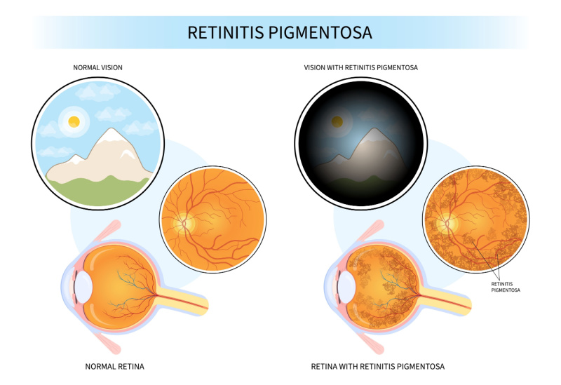
In the typical progression of RP, rod cells begin to malfunction or die off early in the disease process. This leads to the hallmark symptom of nyctalopia, or night blindness. As the rod cells in the mid-peripheral retina succumb, the patient experiences a narrowing of the visual field, often referred to as “tunnel vision.” Over time, the degeneration spreads inward toward the macula—the center of the retina responsible for sharp, detailed, color vision. This secondary involvement of cone cells accounts for the later stages of the disease, where central visual acuity is compromised and color perception becomes distorted.
Genetic Complexity and Inheritance
RP is not caused by a single gene mutation; rather, it is a clinical manifestation of hundreds of different genetic defects. Research has identified mutations in over 100 genes that can lead to an RP phenotype. These mutations affect various cellular processes, including phototransduction—the pathway by which light is converted into vision—as well as protein folding, structural integrity of the outer segments, and intracellular transport.
The inheritance patterns for RP are equally diverse:
- Autosomal Recessive: This is the most common form, where a child inherits one mutated gene from each parent.
- Autosomal Dominant: In this pattern, an individual only needs one copy of the mutated gene from one parent to manifest the condition.
- X-linked: This form, predominantly affecting males, occurs when the mutation is located on the X chromosome.
- Simplex/Sporadic: Many cases arise in individuals with no prior family history, often due to de novo mutations.
Clinical Diagnosis and Staging
Diagnosing retinitis pigmentosa requires a comprehensive ophthalmic evaluation. Because early symptoms may be subtle or mistaken for other vision issues, clinical tools are essential for confirming the diagnosis and monitoring the rate of progression.
Standardized Diagnostic Testing
Ophthalmologists typically use a combination of structural and functional tests to identify the signature markers of RP.
- Fundus Examination: Using an ophthalmoscope, doctors look for the classic “triad” of RP: bone-spicule pigmentary deposits, attenuation of the retinal blood vessels, and a waxy, pale appearance of the optic nerve head.
- Electroretinography (ERG): This is the gold standard for diagnosis. An ERG measures the electrical response of the retina to light. In RP patients, the ERG signal is often severely reduced or completely flat, indicating widespread photoreceptor dysfunction.
- Visual Field Testing: Perimetry is used to map the extent of the patient’s field of vision. This helps track the narrowing of the periphery over time.
- Optical Coherence Tomography (OCT): OCT provides cross-sectional imaging of the retina, allowing clinicians to measure the thickness of retinal layers and track the progressive thinning of the outer nuclear layer where photoreceptor cell bodies reside.
Genetic Testing and Counseling
Given the genetic variety of RP, genetic testing is increasingly standard practice. Identifying the specific causative mutation is not only important for prognosis but is also a prerequisite for accessing emerging gene-specific therapies. Genetic counseling provides families with the information needed to understand recurrence risks and prepare for the future.
The Evolving Landscape of Management and Treatment
Historically, a diagnosis of retinitis pigmentosa carried an atmosphere of hopelessness, with limited options for intervention. Today, however, the medical community is moving toward a more proactive, personalized approach. While there is no current “cure” that reverses the damage, research is advancing rapidly.
Supportive Care and Lifestyle Adaptation
Managing RP is currently focused on maximizing remaining vision and maintaining quality of life.
- Low Vision Rehabilitation: Occupational therapists and low-vision specialists help individuals navigate daily tasks using assistive technology, magnification devices, and orientation and mobility training.
- Nutritional Supplements: Some studies suggest that high-dose Vitamin A palmitate may slow the decline of retinal function in certain patients, though this remains a subject of ongoing clinical debate and should only be undertaken under strict medical supervision due to potential side effects.
- Blue Light Protection: Many specialists recommend wearing sunglasses with high-quality UV and blue-light protection to mitigate potential light-induced stress on compromised retinal cells.
Emerging Therapies: Gene Editing and Beyond
The future of RP treatment lies in molecular medicine.
- Gene Therapy: The most significant breakthrough in recent years is the development of gene-replacement therapies, such as the FDA-approved treatment for mutations in the RPE65 gene. By introducing a functional copy of the gene into the retinal cells, scientists are successfully restoring vision in patients who were previously legally blind.
- Retinal Prosthetics: For patients who have reached the end stage of the disease where no photoreceptors remain, visual implants, or “bionic eyes,” provide a pathway for functional sight. These devices capture light via a camera and transmit signals to an electrode array implanted on the retina, stimulating the remaining neural circuitry to provide a basic perception of light and shapes.
- Optogenetics: This experimental field involves modifying non-photoreceptive cells in the retina to become light-sensitive. By introducing light-sensitive proteins, researchers hope to essentially turn other retinal cells into a surrogate for the lost photoreceptors.
Living with Retinitis Pigmentosa
The psychological and practical implications of RP are significant. Because the disease is progressive and often manifests during the prime working years of an individual’s life, it requires profound adjustments in both professional and personal spheres.
The Importance of Early Intervention
Early diagnosis is crucial, not just for monitoring health but for early involvement with support networks. Connecting with organizations dedicated to retinal degenerative diseases can provide invaluable resources for mental health support, legal advocacy, and community building.

The Future Outlook
The pace of scientific discovery in the field of retinal dystrophies is unprecedented. As we transition from understanding the genetic roadmap of RP to deploying highly specific genomic therapies, the potential to halt, or even reverse, this condition is no longer a matter of “if,” but “when.” The combination of diagnostic precision, advancements in ocular imaging, and the promise of gene therapy and regenerative medicine ensures that the narrative surrounding retinitis pigmentosa is shifting from one of inevitable darkness to a landscape defined by hope, innovation, and technological resilience.
Individuals living with RP remain central to this progress. Their participation in clinical trials and natural history studies continues to provide the data necessary to refine treatments, ensuring that the legacy of this condition will eventually be defined by the successful methods developed to overcome it.
aViewFromTheCave is a participant in the Amazon Services LLC Associates Program, an affiliate advertising program designed to provide a means for sites to earn advertising fees by advertising and linking to Amazon.com. Amazon, the Amazon logo, AmazonSupply, and the AmazonSupply logo are trademarks of Amazon.com, Inc. or its affiliates. As an Amazon Associate we earn affiliate commissions from qualifying purchases.