
The human eye, a marvel of biological engineering, is also a delicate and complex organ. When anomalies occur within its intricate structures, such as the presence of a gas bubble, the ability to accurately visualize and understand these foreign elements becomes paramount for diagnosis and treatment. For a patient, the experience of a gas bubble in the eye can be disorienting and alarming, often described as a shimmering, mobile dark sphere or a cluster of smaller bubbles, which can distort vision dramatically. However, for the medical professional, simply understanding the patient’s subjective experience is insufficient. Modern ophthalmology relies heavily on a sophisticated arsenal of technology to objectively “see” what’s inside, precisely characterize the bubble, and monitor its behavior. This exploration delves into the technological innovations that transform a vague patient description into a detailed, actionable visual understanding.
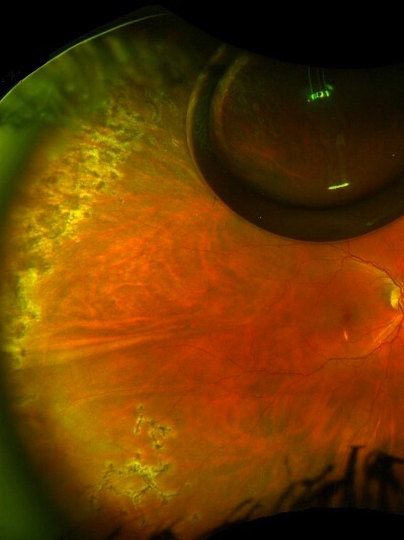
The Unseen World: Bridging the Gap Between Perception and Precision
For centuries, understanding ocular conditions was largely limited to direct observation of the external eye and inferences drawn from patient symptoms. The interior of the eye remained a largely unseen world, its delicate structures obscured by its very design. The question, “what does a gas bubble look like in the eye?” is deceptively simple, encompassing not only the direct physical appearance but also the intricate methods by which we scientifically observe and analyze it.
The Medical Context: When Gas Bubbles Appear
Gas bubbles are not a natural occurrence within the vitreous humor (the jelly-like substance filling the eye). Their presence almost invariably signals a medical intervention or, in rare cases, a traumatic injury. The most common scenario involves vitrectomy surgery, a procedure performed to address retinal detachment, macular holes, or severe vitreous hemorrhage. During vitrectomy, the surgeon may inject a gas bubble (sulfur hexafluoride – SF6, or perfluoropropane – C3F8) into the eye to help reattach the retina or flatten a macular hole. This bubble acts as a temporary internal scaffold, allowing the retina to heal against the back wall of the eye. As the eye’s natural fluid replaces the gas, the bubble slowly diminishes over days or weeks. Other, less common instances include penetrating eye injuries that introduce air, or very rarely, gas-producing infections.
From the patient’s perspective, the gas bubble initially appears as a large, dark, mobile sphere that blocks a significant portion of their vision. As the gas is absorbed and replaced by aqueous humor, the bubble shrinks, often breaking into smaller bubbles, and moves more freely. It can appear silvery, clear, or black, depending on lighting and its position relative to the pupil. The most striking characteristic is its dynamic nature; it shifts with eye movement, creating a constantly changing visual field. The gas-fluid interface is often described as a meniscus or “oil bubble” effect, due to the different refractive indices of gas and fluid, causing light to bend in unusual ways, leading to distortions or a shimmering effect.
Limitations of Traditional Examination
While a skilled ophthalmologist can often detect a large gas bubble with a direct ophthalmoscope, the limitations are significant. Traditional methods offer a two-dimensional view, lack depth perception, and struggle to accurately measure the bubble’s volume, its precise location relative to critical structures (like the retina), or detect smaller associated pathologies. Furthermore, the patient’s experience is subjective. What looks like a “fishbowl” or a “bubble level” to them needs to be translated into objective clinical data. This necessitates technological augmentation, moving beyond the confines of manual observation to a realm of digital precision.
Illuminating the Interior: Breakthroughs in Ocular Imaging Tech
The technological revolution in medical imaging has transformed ophthalmology, offering unprecedented views into the eye’s deepest recesses. These tools allow doctors to not only confirm the presence of a gas bubble but also to meticulously analyze its characteristics and monitor the surrounding tissues.
Optical Coherence Tomography (OCT): A Deeper Dive
Perhaps the most transformative technology in modern ophthalmology is Optical Coherence Tomography (OCT). OCT is a non-invasive imaging test that uses light waves to take cross-section pictures of your retina. Think of it as an optical ultrasound, but instead of sound waves, it uses light. This technology provides incredibly high-resolution, cross-sectional images of the retinal layers and the vitreous, revealing structures invisible to the naked eye or even through traditional fundus photography.
When a gas bubble is present, OCT reveals it as a black, optically empty space within the vitreous cavity. The distinct interface between the gas and the surrounding vitreous fluid or retina is sharply delineated. OCT can precisely measure the bubble’s dimensions, track its shrinkage over time, and, crucially, assess the integrity of the underlying retina. For instance, in a patient recovering from retinal detachment surgery, OCT can confirm that the gas bubble is effectively tamponading (pushing against) the retina, ensuring it remains attached. It can also detect subtle fluid accumulation beneath the retina or the development of epiretinal membranes, which might be obscured by the gas bubble itself in less advanced imaging. The ability to generate 3D OCT scans further enhances understanding, allowing clinicians to rotate and analyze the bubble’s spatial relationship to surrounding structures.
Ultrasound Biomicroscopy (UBM) and Its Applications
While OCT excels at imaging the posterior segment of the eye, particularly the retina, other technologies are vital for different regions. Ultrasound Biomicroscopy (UBM) uses high-frequency ultrasound to create detailed images of the anterior segment of the eye (cornea, iris, ciliary body, and lens) and structures immediately behind the lens that might be difficult to visualize with light-based technologies due to opacity (e.g., dense cataracts).
Though less frequently used for posterior vitreous gas bubbles than OCT, UBM can be invaluable in specific scenarios. For instance, if a gas bubble is located in the anterior vitreous and the view is obstructed by significant corneal edema or other anterior segment opacities, UBM can penetrate these barriers to visualize the bubble. It works by sending sound waves into the eye and capturing the echoes, which are then converted into an image. Gas, being a poor conductor of sound, would appear as a highly reflective surface, often with a “shadowing” effect behind it, indicating the presence of gas. Its role is often complementary, particularly when light-based imaging is compromised.
Fundus Photography and Angiography: Capturing the Surface and Beyond
Fundus photography involves taking a high-resolution photograph of the fundus, which includes the retina, optic disc, macula, and posterior pole. While it provides a wide-field view, its primary limitation when a gas bubble is present is that the bubble itself often obscures parts of the fundus, creating a dark, featureless area in the photograph. However, as the bubble shrinks, fundus photography can be used to document the overall health of the retina, detect any residual issues, and monitor the resolution of the gas.
Fluorescein angiography, a more specialized form of fundus photography, involves injecting a fluorescent dye into the bloodstream. The dye travels to the blood vessels in the eye, and a special camera takes pictures as the dye illuminates these vessels. While gas bubbles do not directly appear on angiography, this technique is crucial for assessing retinal circulation and detecting any leakage or abnormal vessel growth that might be associated with the underlying condition that necessitated the gas injection, or any complications arising from it. The gas bubble might still cast a shadow, but the angiography can provide vital information about the perfused retina around and beyond the bubble’s immediate obstruction.

Beyond Static Images: Dynamic Visualization and Simulation
The ability to capture static images is a monumental leap, but the future of ocular diagnostics and training involves dynamic and immersive experiences. These technologies push the boundaries of “what it looks like” by allowing interaction, simulation, and predictive modeling.
Virtual Reality (VR) and Augmented Reality (AR) in Ophthalmology Training
For aspiring ophthalmologists, understanding the three-dimensional anatomy of the eye and the implications of procedures like vitrectomy can be challenging. VR and AR technologies are revolutionizing surgical training by creating highly realistic simulations. In a VR environment, trainees can “perform” vitrectomies, inject gas bubbles, and observe their behavior in a simulated eye, learning how the bubble moves, settles, and impacts the visual field. This allows them to virtually experience “what a gas bubble looks like” from the patient’s perspective and from the surgeon’s operating microscope.
AR, on the other hand, can overlay digital information onto a real-world view. During live surgery, AR could potentially project real-time OCT data onto the surgeon’s view, allowing them to see the exact relationship between their instruments, the gas bubble, and delicate retinal structures simultaneously, enhancing precision and safety. This blends the physical and digital, providing an enriched visual understanding that static images alone cannot offer.
AI-Powered Diagnostics and Predictive Modeling
Artificial Intelligence (AI) is rapidly becoming an indispensable tool in medical imaging and diagnostics. AI algorithms can be trained on vast datasets of OCT scans, fundus photos, and patient data to identify patterns that might be imperceptible to the human eye. For a gas bubble in the eye, AI can:
-
Automated Bubble Detection and Measurement: AI can automatically detect the presence of gas bubbles, measure their volume, and track their shrinkage rate with high precision, freeing up clinicians’ time.
-
Predictive Modeling of Retinal Reattachment: By analyzing the characteristics of the gas bubble and the underlying retinal condition, AI models could potentially predict the likelihood of successful retinal reattachment or identify patients at higher risk of complications.
-
Early Detection of Complications: AI can flag subtle changes in retinal morphology around the gas bubble that might indicate impending issues like inflammation or membrane formation, allowing for earlier intervention.
This predictive and analytical power transforms “what it looks like” into “what it means” and “what might happen next,” moving beyond pure observation to proactive management.
The Role of 3D Reconstruction in Patient Education
While clinicians have access to sophisticated imaging, explaining the presence and behavior of an intraocular gas bubble to a patient can be challenging. Static 2D images often fail to convey the dynamic, volumetric reality. 3D reconstruction technology, built from a series of 2D OCT or ultrasound images, creates a realistic, rotatable 3D model of the eye’s interior, including the gas bubble.
This technology is invaluable for patient education. Patients can visually grasp the size, shape, and position of their gas bubble, understanding why it affects their vision in a particular way and how it will gradually disappear. Seeing a personalized 3D model can significantly reduce anxiety and improve adherence to post-operative instructions, as patients have a clearer understanding of their own unique condition.
The Future of Ocular Visualization: Enhancing Patient Outcomes
The trajectory of technological advancement in ophthalmology points towards even more integrated, personalized, and accessible methods of visualizing and understanding ocular conditions, including the elusive gas bubble.
Miniaturization and Portability of Imaging Devices
Currently, high-resolution imaging devices like OCT machines are relatively large and expensive. The future promises miniaturized, portable, and even handheld devices that can deliver comparable imaging quality. This would allow for more frequent monitoring in various settings, including outpatient clinics, remote locations, and even potentially for home use, enabling patients to track their bubble’s resolution under telemedical guidance. Such advancements would democratize access to advanced diagnostics, further enhancing the understanding of “what a gas bubble looks like” for a wider population.
AI-Driven Personalization of Treatment
As AI capabilities grow, they will move beyond diagnosis to offer personalized treatment recommendations. For patients with intraocular gas bubbles, this could mean AI-optimized post-operative positioning advice, tailored medication regimens, or even predicting the ideal gas composition and volume for individual surgical cases to maximize success rates and minimize recovery time. The “look” of the bubble, meticulously analyzed by AI, would directly inform bespoke treatment plans.

Teleophthalmology and Remote Diagnostics
The synergy of portable imaging devices, AI analysis, and robust telecommunications infrastructure will accelerate the adoption of teleophthalmology. Patients with gas bubbles could have their eyes imaged remotely, with the data transmitted to specialists for AI-assisted analysis and interpretation. This would be particularly beneficial for follow-up care, reducing the need for frequent in-person clinic visits and making expert care accessible regardless of geographical barriers. The remote visualization of a gas bubble’s status would become a routine part of post-operative management, transforming how patients and doctors perceive and manage this specific ocular phenomenon.
In conclusion, “what does a gas bubble look like in the eye” is a question whose answer is increasingly shaped by the remarkable progress in medical technology. From the intricate cross-sections provided by OCT to the immersive training offered by VR, and the predictive power of AI, technology not only allows us to see these transient entities with unparalleled clarity but also to understand their implications, predict their behavior, and ultimately, enhance patient care and outcomes. The journey from a patient’s blurry perception to a clinician’s precise diagnosis is a testament to the ingenuity of technological innovation in ophthalmology.
aViewFromTheCave is a participant in the Amazon Services LLC Associates Program, an affiliate advertising program designed to provide a means for sites to earn advertising fees by advertising and linking to Amazon.com. Amazon, the Amazon logo, AmazonSupply, and the AmazonSupply logo are trademarks of Amazon.com, Inc. or its affiliates. As an Amazon Associate we earn affiliate commissions from qualifying purchases.