
The landscape of modern medicine is being fundamentally reshaped by the integration of sophisticated technology, shifting the traditional laboratory experience from a manual, labor-intensive process to a streamlined, digital-first operation. When we ask “what are epithelial cells in the urine,” we are no longer just looking through the lens of biological inquiry; we are looking at a complex data point within the burgeoning field of HealthTech. In the digital age, the identification and quantification of these cells—which line the surfaces of the body—represent a triumph of computer vision, machine learning, and automated diagnostic hardware.
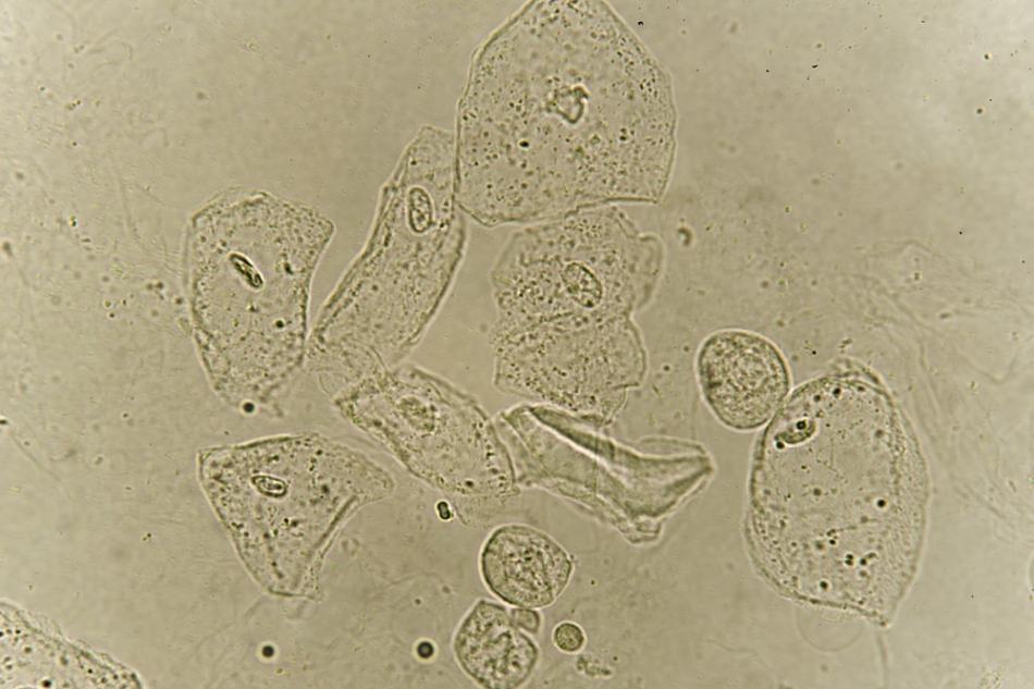
The shift toward automated urinalysis has revolutionized how clinics and hospitals process patient samples. Understanding the presence of squamous, transitional, or renal tubular epithelial cells is now a task managed by high-speed sensors and neural networks. This technological evolution ensures higher precision, faster turnaround times, and a scalable approach to population health management.
The Evolution of Urinalysis: From Manual Microscopy to Automated Recognition Systems
For decades, the standard method for identifying epithelial cells involved a technician manually peering through a microscope. This process was prone to human error, subjective interpretation, and significant time lag. Today, the tech industry has replaced the manual slide with automated sediment analyzers that utilize two primary technological frameworks: Flow Cytometry and Digital Image Recognition.
Automated Flow Cytometry in Liquid Biopsy
Automated flow cytometry utilizes laser technology to analyze cells as they flow in a single file through a sensing zone. In the context of detecting epithelial cells, the hardware measures the light scatter and fluorescence of each particle. By applying specific algorithms, the system can differentiate between cell types based on their size, granularity, and complexity. This “liquid biopsy” tech allows for the processing of hundreds of samples per hour, a feat impossible for human technicians.
High-Resolution Digital Imaging and Computer Vision
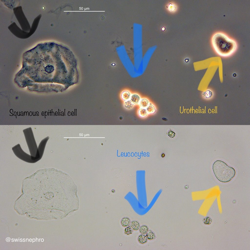
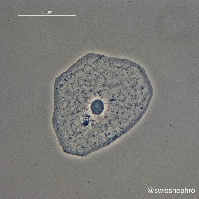
The most significant leap in diagnostic tech is the use of high-resolution digital imaging. Modern analyzers capture thousands of digital frames of a urine sample. Software then segments these images, isolating individual particles. Computer vision models, specifically designed for urological diagnostics, compare these isolated images against a massive database of verified cellular structures. This tech does not just see a cell; it categorizes it as a squamous epithelial cell (often a sign of sample contamination) or a renal tubular cell (a potential indicator of kidney pathology) with surgical precision.
The Role of Artificial Intelligence and Deep Learning in Cellular Classification
The true “brain” behind modern urinalysis is the suite of Artificial Intelligence (AI) tools that interpret raw data. Deep learning models, particularly Convolutional Neural Networks (CNNs), have become the gold standard for classifying epithelial cells in a digital environment.
Neural Network Training and Pattern Recognition
To reach a high level of accuracy, these AI models are trained on millions of labeled images. By processing these datasets, the software learns the subtle morphological differences between cell types. For example, the software can distinguish the large, flat appearance of squamous cells from the smaller, rounder profile of renal tubular cells. This level of pattern recognition reduces the “noise” in diagnostic data, allowing clinicians to focus on high-priority results flagged by the system.
Predictive Analytics and Trend Mapping
Beyond simple identification, AI-driven platforms are now capable of predictive analytics. By integrating a patient’s historical urine data into a cloud-based dashboard, software can identify trends. If the concentration of renal epithelial cells shows a marginal but steady increase over six months, the system can trigger an automated alert. This proactive approach to health tech moves the needle from reactive treatment to preventive maintenance, powered entirely by data science.
Reducing Inter-observer Variability
One of the greatest challenges in traditional pathology is inter-observer variability—the reality that two experts might interpret the same slide differently. Software eliminates this inconsistency. By utilizing a standardized algorithmic approach, a diagnostic lab in New York and a clinic in Singapore can achieve identical results for the same sample, ensuring a global standard of care driven by code rather than subjective human sight.
Telehealth Integration and the Consumerization of Diagnostic Data
The technology surrounding the detection of epithelial cells has moved out of the laboratory and into the palms of patients’ hands. The “consumerization” of healthcare tech has led to the development of patient portals and smartphone-linked diagnostic tools that interpret complex lab results for the end-user.
Smart Portals and Natural Language Processing
When a patient receives a report mentioning “epithelial cells,” their first instinct is to seek clarity. Modern Electronic Health Record (EHR) systems now utilize Natural Language Processing (NLP) to translate cryptic lab values into actionable insights. These portals provide interactive visualizations, showing where a patient’s levels sit on a digital spectrum and providing context-aware explanations of what those technological readings mean for their specific health profile.
The Rise of At-Home Testing and IoT
We are witnessing the emergence of the “Smart Toilet” and smartphone-based dipstick readers. These Internet of Things (IoT) devices use multi-spectral imaging to analyze urine samples at home. While currently more focused on chemical markers (like pH or glucose), the roadmap for these devices includes miniaturized optical sensors capable of detecting cellular presence. This decentralized approach to testing relies on secure cloud synchronization, where data is sent from a home device to a physician’s dashboard in real-time.
APIs and Ecosystem Interoperability
The modern health tech stack relies on Interoperability. Application Programming Interfaces (APIs) allow different pieces of software—the lab’s analyzer, the hospital’s record system, and the patient’s fitness app—to communicate. When epithelial cell counts are logged, this data can be cross-referenced with other digital metrics like hydration levels or medication logs, providing a holistic view of the user’s biological state.
Cybersecurity and Ethical Considerations in Digital Health Mapping
As the identification of epithelial cells becomes a purely digital process, the security of that data becomes paramount. The transition from physical slides to digital files introduces new risks that the tech industry must address through robust cybersecurity frameworks.
Data Encryption and HIPAA Compliance
Every digital image of a cellular sediment is protected health information (PHI). Tech providers in this space must utilize end-to-end encryption to ensure that data moving from an automated analyzer to the cloud remains uncompromised. Compliance with standards like HIPAA in the US or GDPR in Europe is baked into the software architecture, ensuring that the digitizing of urological health does not come at the cost of personal privacy.
The Ethics of AI Decision-Making
As we rely more on software to identify epithelial cells, the question of “algorithmic transparency” arises. It is vital for tech developers to ensure their AI models are free from bias and that their decision-making processes are auditable. This “Explainable AI” (XAI) ensures that if a software flags a sample as high-risk, a human clinician can trace the logic back to the specific pixels and patterns the AI identified.
Blockchain for Diagnostic Integrity
Some emerging health tech platforms are exploring blockchain technology to create an immutable record of diagnostic results. By anchoring lab findings—including cell counts and types—to a decentralized ledger, patients and providers can ensure the integrity of the data over time. This prevents unauthorized tampering and provides a “single source of truth” for a patient’s long-term diagnostic history.
The technological infrastructure required to answer “what are epithelial cells in the urine” is a testament to the power of the modern digital stack. From the hardware that captures the image to the AI that interprets the pixels and the secure cloud that stores the result, the process is a masterclass in how technology is refining our understanding of the human body. As these tools continue to evolve, the speed, accuracy, and accessibility of diagnostic data will only increase, marking a new era of data-driven wellness.
aViewFromTheCave is a participant in the Amazon Services LLC Associates Program, an affiliate advertising program designed to provide a means for sites to earn advertising fees by advertising and linking to Amazon.com. Amazon, the Amazon logo, AmazonSupply, and the AmazonSupply logo are trademarks of Amazon.com, Inc. or its affiliates. As an Amazon Associate we earn affiliate commissions from qualifying purchases.